Figure 4. Immunofluorescence staining for α-SMA in fibrotic healing corneas at different time points. A: Control cornea exhibited no α-SMA staining. B: At 3 days after injury, no α-SMA staining was observed. C: At 1 week after injury, α-SMA had appeared in the most anterior stroma. D: At 2 weeks after injury, α-SMA had appeared in the entire repairing stroma. E: At 3 weeks after injury, α-SMA had appeared in the most posterior stroma. F-G: At 1 to 2 months after injury, α-SMA was detected in the posterior stroma, and the positively stained area for α-SMA had
reduced. H: At 3 months after injury, α-SMA staining was nearly absent in the wound stroma. Green staining indicates α-SMA protein.
Blue is DAPI staining of cell nuclei. (E) is the epithelium, and (S) is the stroma. Magnification: ×100. Scale bar: 250 μm.
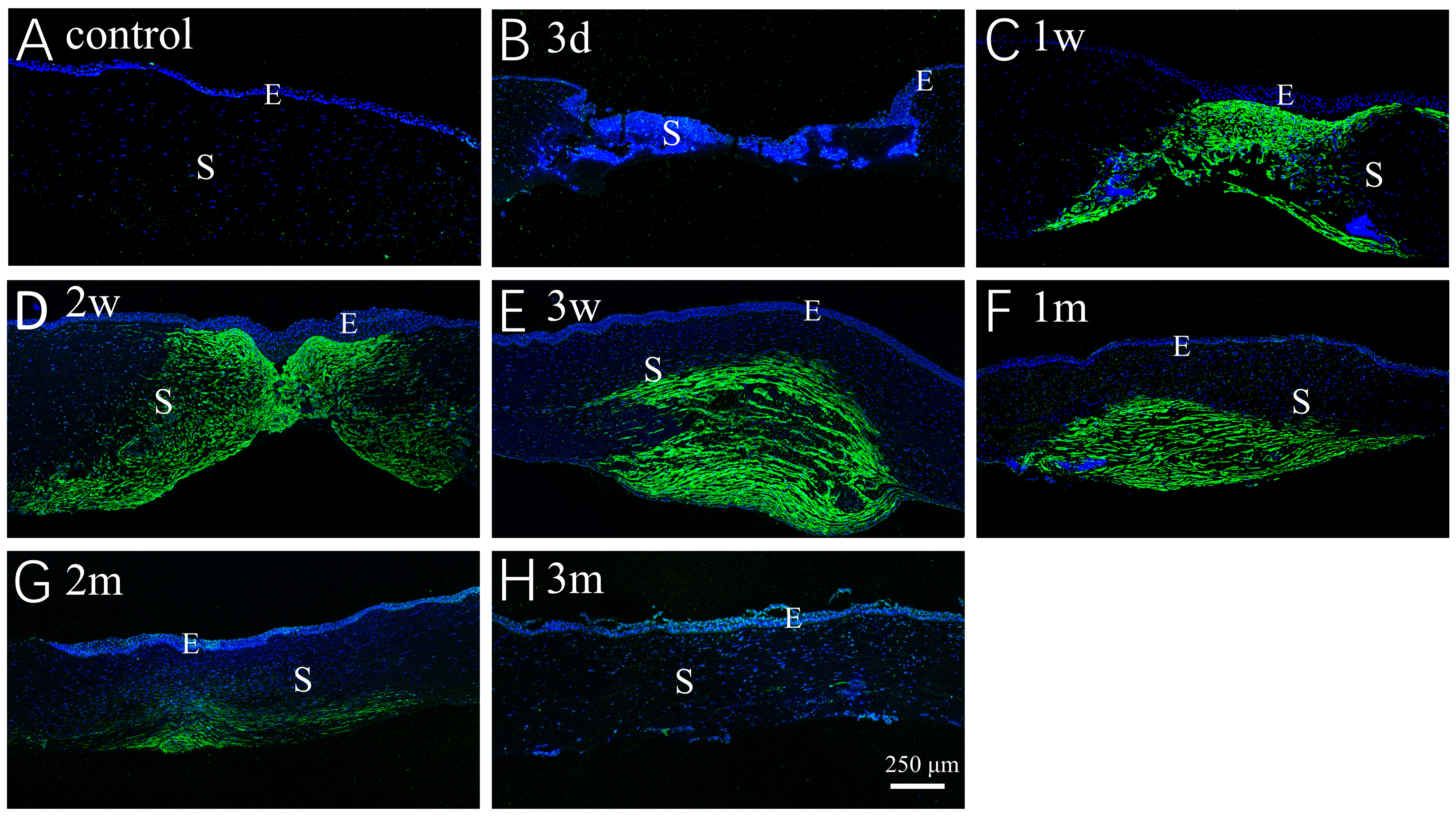
 Figure 4 of
Meng, Mol Vis 2023; 29:58-67.
Figure 4 of
Meng, Mol Vis 2023; 29:58-67.