Figure 3. Immunofluorescence staining for TGF-β1 in two different corneas at each time point. In the normal cornea, no TGF-β1 protein
was detected in the stroma. At 3 days after injury, a large quantity of TGF-β1 was deposited in the acellular fibrin clot.
At 1 week after injury, TGF-β1 was localized to the entire healing stroma. At 2 weeks after injury, the deposition of TGF-β1
had decreased in the anterior stroma, and TGF-β1 was localized to the posterior stroma. At 3 weeks after injury, TGF-β1 was
nearly absent in the anterior stroma, and TGF-β1 had decreased in the posterior stroma. At 1 month after injury, TGF-β1 had
further decreased, and TGF-β1 was only weakly detected in the posterior stroma. At 2 and 3 months after injury, TGF-β1 was
absent in the repairing stroma. Green staining indicates TGF-β1 protein. Blue is DAPI staining of cell nuclei. (E) is the
epithelium. (S) is the stroma. Magnification: ×100. Scale bar: 250 μm.
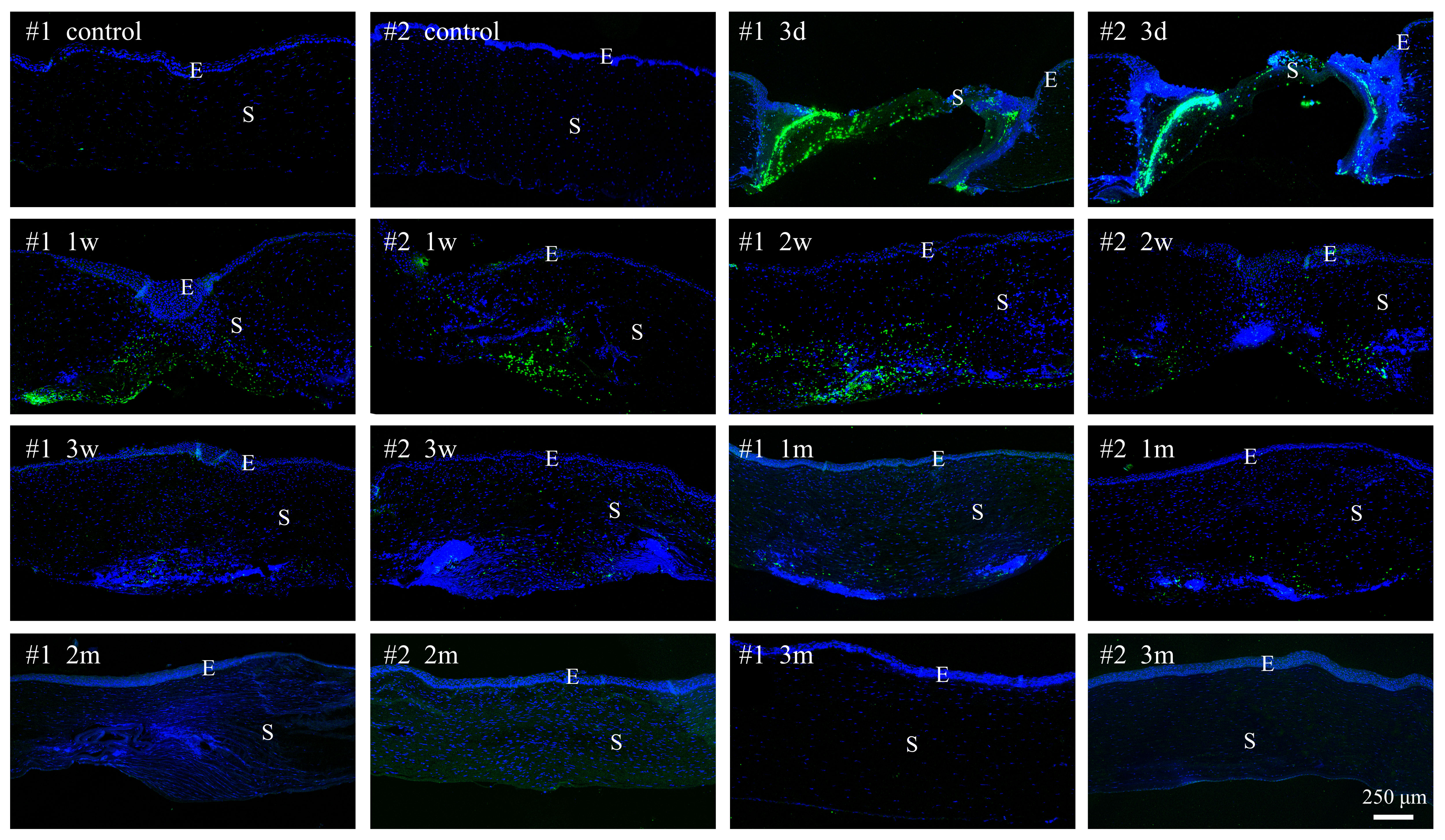
 Figure 3 of
Meng, Mol Vis 2023; 29:58-67.
Figure 3 of
Meng, Mol Vis 2023; 29:58-67.