Figure 1. Corneal haze of rabbits. A: Slit lamp photographs of rabbit corneal opacity at different time points. Normal unwounded cornea was transparent. Three
days after injury, the wound area was filled with translucent fibrous plaques. Fine iris and lens details could be observed
1 week after injury, a prominent haze appeared in the wound margin, and details of the iris could be observed to some extent.
Two weeks after injury, increased haze appeared in the wound margin, with mild obscuration of the iris details. Three weeks
after injury, a dense opacity appeared in the wound area, with moderate obscuration of the iris details and lens. One month
after injury, complete opacification was observed in the wound area, and the iris was not visible. Two months after injury,
a focal transparent area appeared in the wound area. Three months after injury, the iris was faintly visible. B: Changes in corneal opacity scores at different time points. Corneal opacity appeared at 3 days, then gradually increased,
peaking at 1 month before decreasing. The level of corneal haze at each time point after injury was significantly higher than
in the control group. The 1 m group exhibited a significantly higher haze score than other groups. Data are expressed as mean
± SD, n=6.
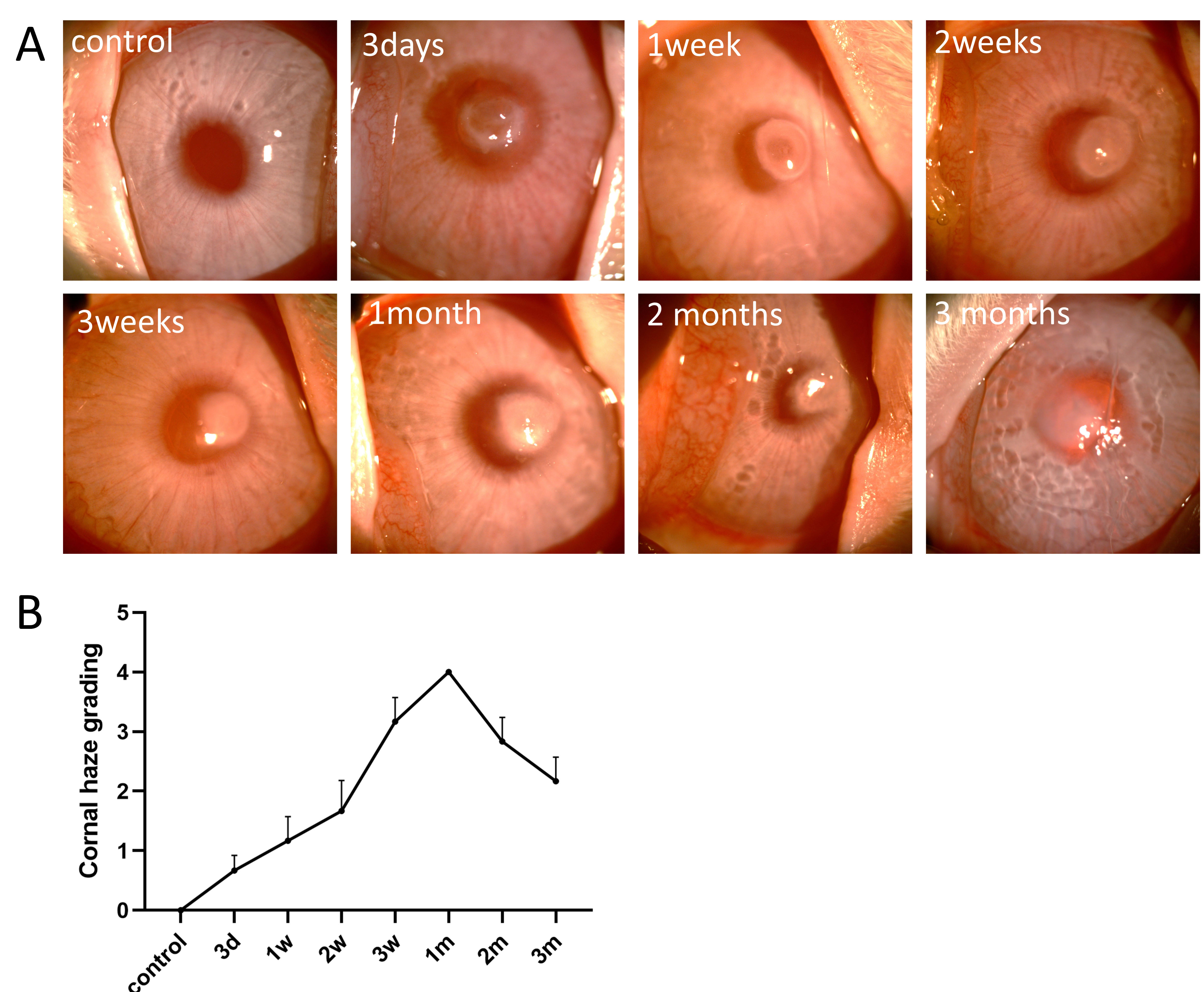
 Figure 1 of
Meng, Mol Vis 2023; 29:58-67.
Figure 1 of
Meng, Mol Vis 2023; 29:58-67.