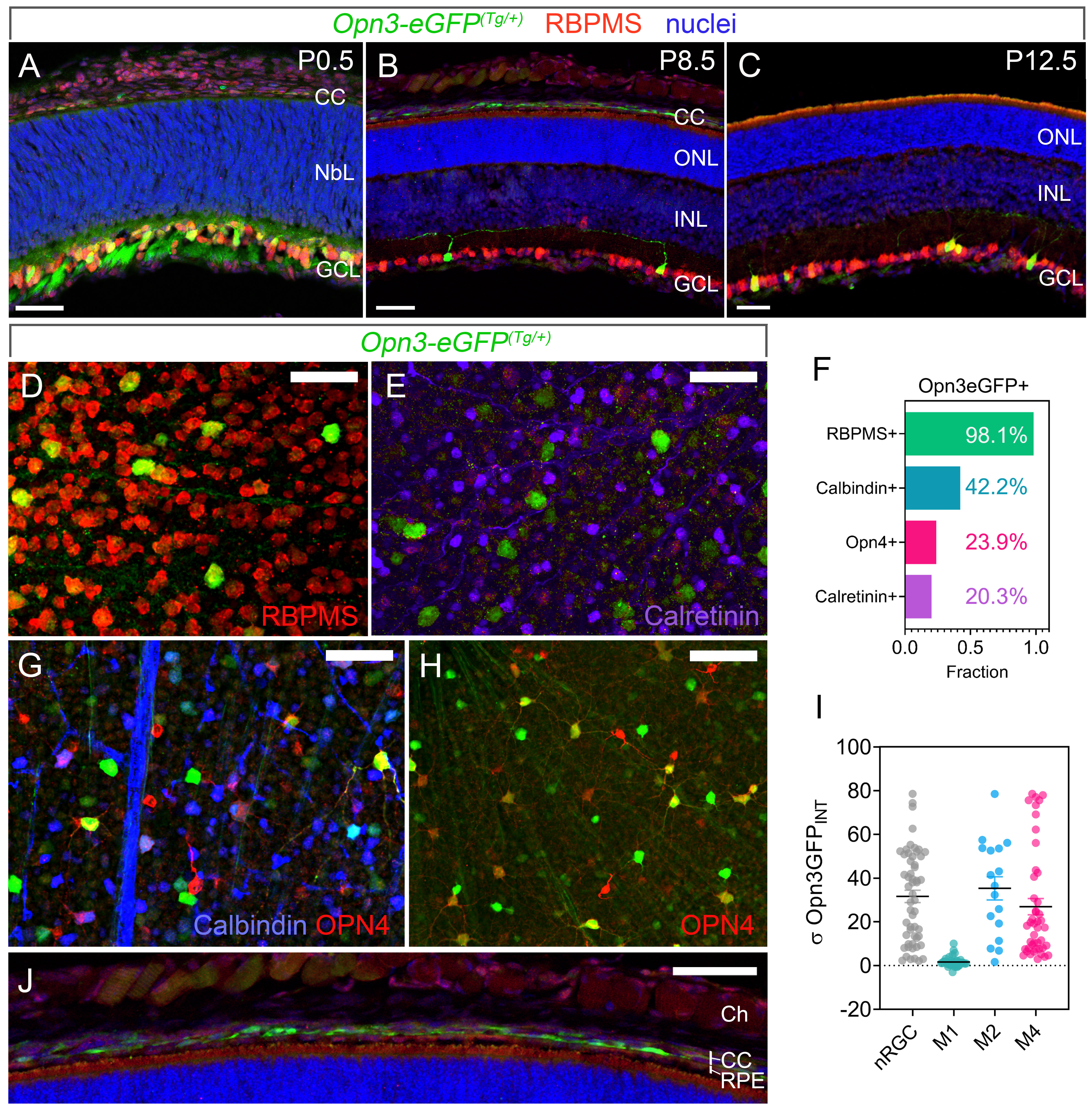
Figure 1. Opn3 is expressed in retinal ganglion cells. A–C: Retinal expression of eGFP (green) in Opn3-eGFP(Tg/+) mice at P0.5 (A), P8.5 (B), and P12.5 (C; 50 µM scale bars), showing expression within RBPMS-labeled (red) retinal ganglion cells (RGCs) in the ganglion cell layer
(GCL). Nuclei are DAPI labeled (blue). The proportion of ganglion cells that express Opn3-eGFP is high at P0.5 (A) and reduced by P8.5 (B). Opn3-eGFP is also expressed in cells of the choriocapillaris (CC; A, B). D, E: Whole-mount retina from Opn3-eGFP(Tg/+) mice at P16 labeled for GFP (green) and melanopsin (red; D) or GFP (green) calbindin (blue) and calretinin (magenta; 25 µM scale bars). F: Chart showing the proportion of Opn3-eGFP(Tg/+) cells that are positive for RBPMS (D), calretinin (E), calbindin (G, scale bar 25 µM), and OPN4 (H, scale bar 50 µM). I: Along with the morphological assessment of dendritic arbors, this analysis identified populations of Opn3-eGFP RGCs that did not express OPN4 (nRGC: gray), the M1 subtype of OPN4 RGC, which did not express Opn3-eGFP (M1-ipRGC: green), and two subtypes of OPN4 RGC, the M2 (M2-ipRGC: blue) and M4 (M4-ipRGC: red), which expressed both OPN4
and Opn3-eGFP. This is represented in the chart showing the relative expression levels of Opn3-eGFP in RGCs that do not express OPN4 (nRGCs) as well as the M1, M2, and M4 OPN4 RGCs. (J) Higher magnification image of the Opn3-eGFP
labeling in the choriocapillaris (CC). NbL: neuroblastic layer. ONL: outer nuclear layer. INL: inner nuclear layer (50 µM
scale bar). RPE: retinal pigment epithelium.

 Figure 1 of
Linne, Mol Vis 2023; 29:39-57.
  Figure 1 of
Linne, Mol Vis 2023; 29:39-57.