Figure 2. Clinical photographs of Case 1 (A-D), Case 2 (E-H), and Case 3 (I-L). Front facial profile and side facial profile of the
patients illustrate the presence of a characteristic geriatric appearance, with alopecia, sparse eyebrows, frontal bossing,
prominent supraorbital ridges, depressed nasal bridge, anteverted nostrils, and a long philtrum. Clinical photograph of the
oral cavity shows impaction of teeth, with a reduction in tooth number. Radiograph of the face (lateral view with open mouth)
reveals a normal number of teeth, confirming pseudoanodontia. Anterior segment photograph of Case1 (B) reveals the presence
of megalocornea with prominent iridocorneal adhesions and keratopathy (blue arrow). Corneal opacities are also seen (black
arrows; magnified inset). AS-OCT image of the same patient (D) confirms the iridocorneal adhesions (white arrow). Central
keratopathy can be seen in the anterior segment photograph of RE of Case 2 (F). An ultrasound biomicroscopy (UBM) of the same
patient revealed central corneal thickening with a thinned out iris and ciliary body (H).
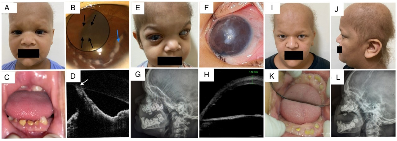
 Figure 2 of
Gupta, Mol Vis 2023; 29:365-377.
Figure 2 of
Gupta, Mol Vis 2023; 29:365-377.