Figure 2. Retinal imaging for subject P2 (A–C). (A) Fundus imaging in P2 demonstrated widespread posterior pole chorioretinal atrophy surrounded by a rim of hyperautofluorescence
(B). (C) SD–OCT imaging revealed widespread retinal atrophy (size bar=200 μm).
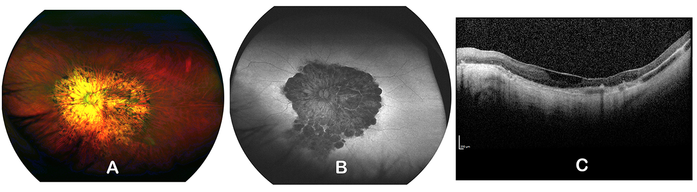
 Figure 2 of
Kolawole, Mol Vis 2023; 29:329-337.
Figure 2 of
Kolawole, Mol Vis 2023; 29:329-337.