Figure 1. Retinal imaging for subject P1 (A–F) compared to a normal subject (G–I). (A, B) Wide-field retinal photography for P1 taken
in 2013 shows pigmentary changes at both the macula plus Pearl degeneration in the peripheral retina. (C) SD–OCT imaging demonstrated an ellipsoid zone deficit (horizontal bar) in the left macula. (D) Repeat retinal photography in 2022 illustrated a dramatic retinal sheen associated with hyperautofluorescence (E) and further enlargement of the ellipsoid zone defect (F). Size bar=200 µm. Detailed assessment of the outer retina (C,F insert, dotted lines) revealed the normal thickness of the outer retina between the outer limiting membrane and the retinal
pigment epithelium (~84 microns), but the absence of stratification delineating the photoreceptor’s outer segments. Small
white arrows indicate (i) the outer limiting membrane; (ii) the ellipsoid zone; (iii) the interdigitating zone; and (iv) Bruch's
membrane/RPE complex. Representative images display the fundus (G), autofluorescence (H), and OCT imaging (I) from an unaffected individual.
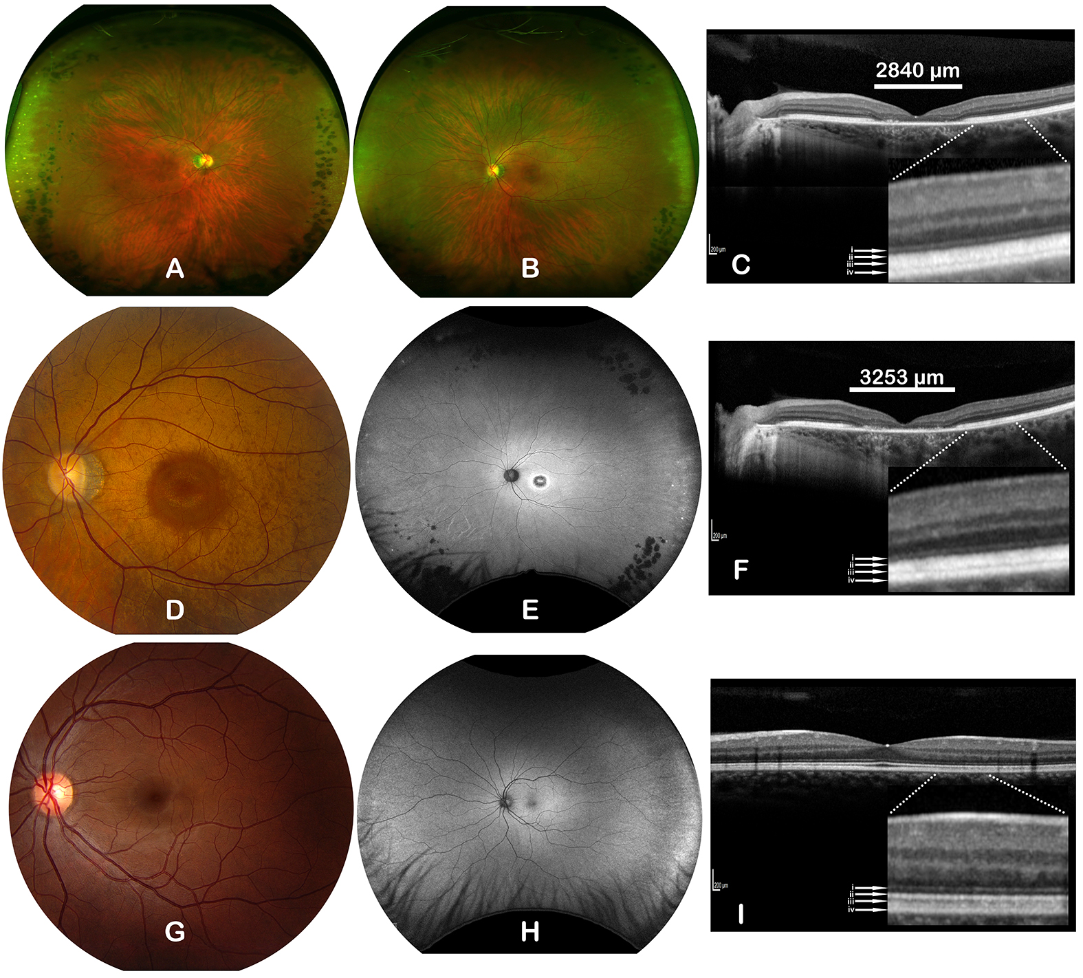
 Figure 1 of
Kolawole, Mol Vis 2023; 29:329-337.
Figure 1 of
Kolawole, Mol Vis 2023; 29:329-337.