Figure 5. SFN suppresses TGF-β-induced MLC phosphorylation in HTFs. A: After a 24-h culture followed by a 24-h starvation, cells were incubated with or without SFN (10 µM) for an additional 24
h. Then, TGF-β (5 ng/ml) was added at indicated time points. Finally, total MLC and phospho-MLC were quantified. Densitometric
analysis of p-MLC/MLC is presented in (B). Data are presented as mean ± SD (n=3). *p<0.05 compared to controls. †p<0.05 compared to cells treated with only TGF-β.
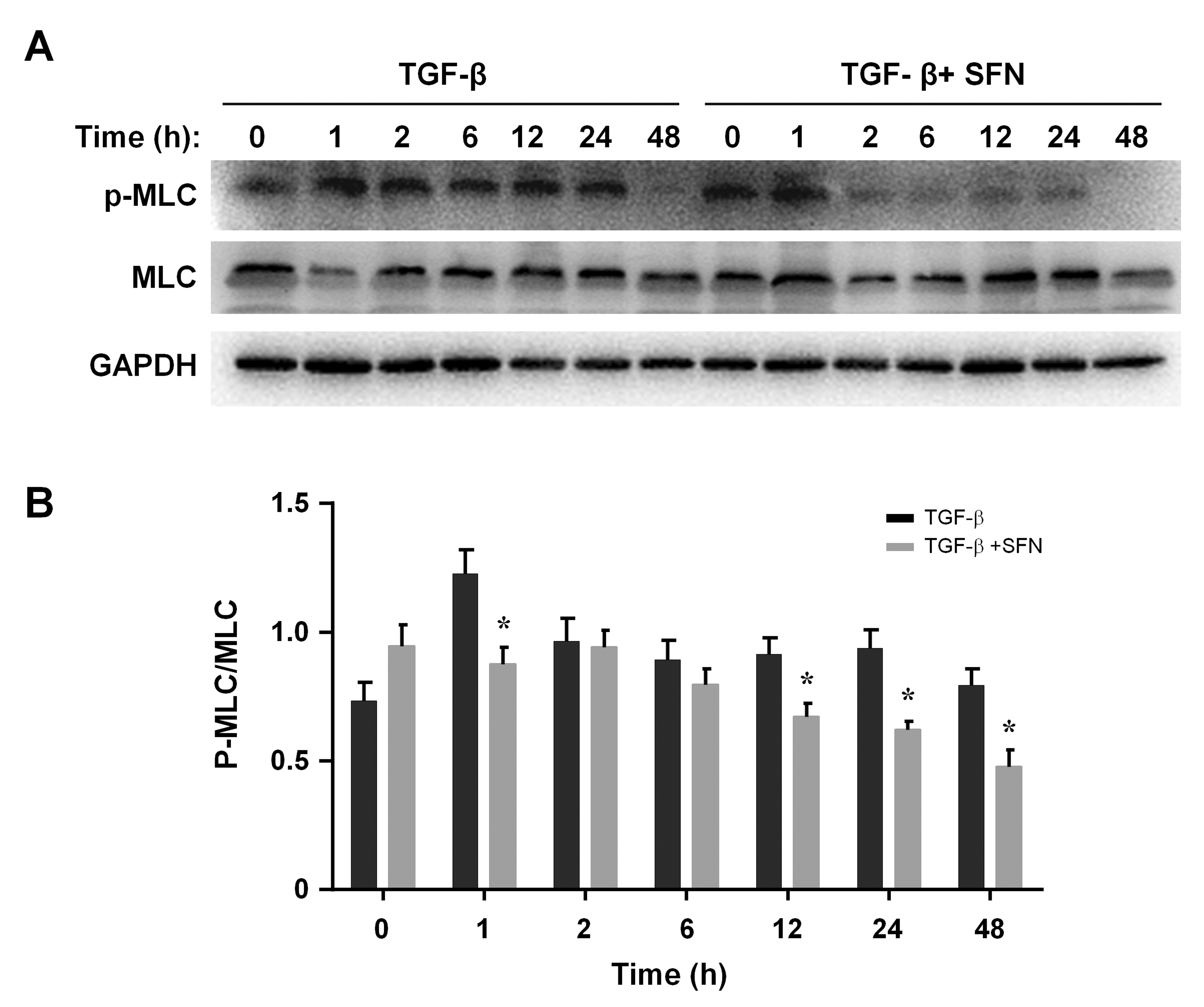
 Figure 5 of
Liu, Mol Vis 2023; 29:306-316.
Figure 5 of
Liu, Mol Vis 2023; 29:306-316.