Figure 5. Mouse corneal epithelium on days 4 (A) and 7 (B and C) after Algerbrush II injury. A: Multiple layers of regenerated conjunctival epithelial cells have accumulated at the limbal region moving toward the corneal
center. B: The limbal epithelium has returned to its normal monocellular layer state 7 days after injury induction (arrowhead). C: The corneal surface is covered by an atrophic monolayer of basal epithelial cells with goblet cells scattered throughout
the surface. Scale bar=50 µm.
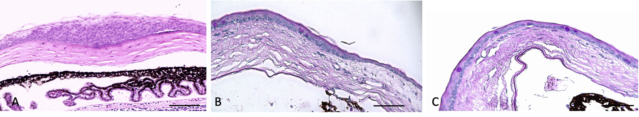
 Figure 5 of
Shadmani, Mol Vis 2023; 29:256-265.
Figure 5 of
Shadmani, Mol Vis 2023; 29:256-265.