Figure 4. Hematoxylin and eosin-stained mouse corneal section visualized under bright light. A: Normal corneal epithelial cells attach to the basement membrane. Corneal spindle shaped keratocytes are distributed throughout
the stromal layer and normal endothelial cells cover the descemet membrane. B: The limbus is a transitional zone where conjunctival epithelial cells transform from monolayered conjunctival epithelial
cells (blue arrow) to multi-layered corneal epithelial cells in the cornea (white arrowhead). The normal anterior chamber
(AC) is clear with no exudate or cells. The normal iris is thin with constricted blood vessels. C: Dilated iris vessels (IV) and exudate deposition on the endothelial layer are illustrated. Longer durations of injury result
in more pigment release into the anterior chamber. D: immediately after Algerbrush injury, the corneal and limbal epithelial cells are absent. [40X and 20X Magnification under
an EVOS XL Core microscope, Scale bar=50 µm]..
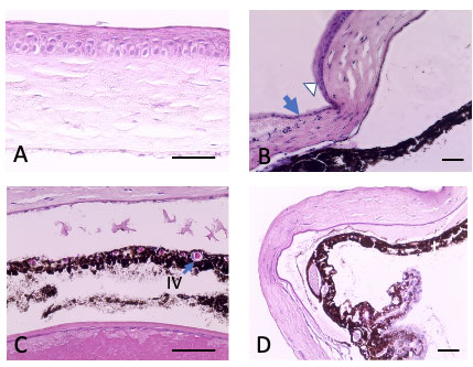
 Figure 4 of
Shadmani, Mol Vis 2023; 29:256-265.
Figure 4 of
Shadmani, Mol Vis 2023; 29:256-265.