Figure 1. Corneal examination immediately after inducing injury with Algerbrush II. Panels A-E show typical images of the eyes before (A) and following injury (B- E) when visualized using slit lamp, fluorescent staining (A.F - E.F) and optical coherence tomography (A.O - E.O). A: Normal, clear cornea and FS pattern with diffuse punctate epithelial defects and normal OCT with clear anterior chamber,
B: Following a 30–45 s injury, the cornea is clear with minimal debris in the anterior chamber, C: 60–75 s injury results in a mild edema of the cornea with mild release of cells and debris into the anterior chamber that
is visible by OCT (C.O), D: A 90–120-s injury results in more edema and the release of cells and pigment into the anterior chamber, confirmed with OCT
images. E: A 3–4 min injury also resulted in significant corneal edema and pigment release. B: The corneal thickness in normal eyes and immediately after injury. Statistical analysis revealed significant differences
between the normal and 60–75 s and 90–120 s injury duration. However, there was no significant difference between the 3–4
min injured and normal group due to development of corneal edema after prolonged injury. (p value: *=0.0200, ***=0.006).
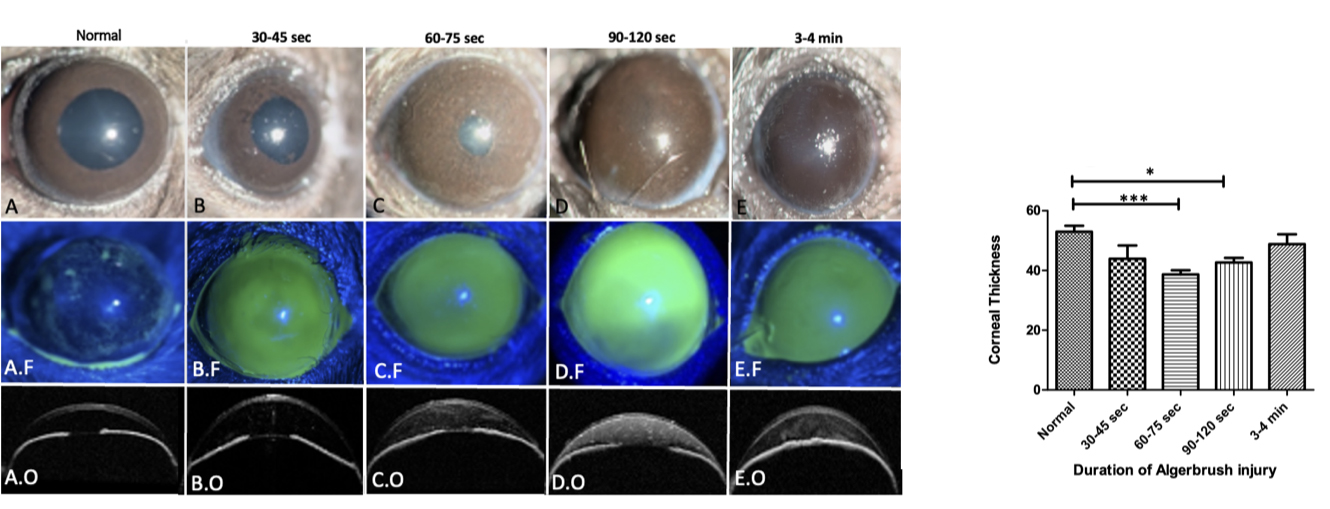
 Figure 1 of
Shadmani, Mol Vis 2023; 29:256-265.
Figure 1 of
Shadmani, Mol Vis 2023; 29:256-265.