Figure 1. Slit-lamp photomicrographs of pre-Descemet corneal dystrophy. Slit-lamp examination of Case 1 (A, right eye), Case 2 (B, right eye), and Case 3 (C, left eye) demonstrating diffuse, punctate, white-gray opacities in the posterior stroma anterior to the Descemet membrane.
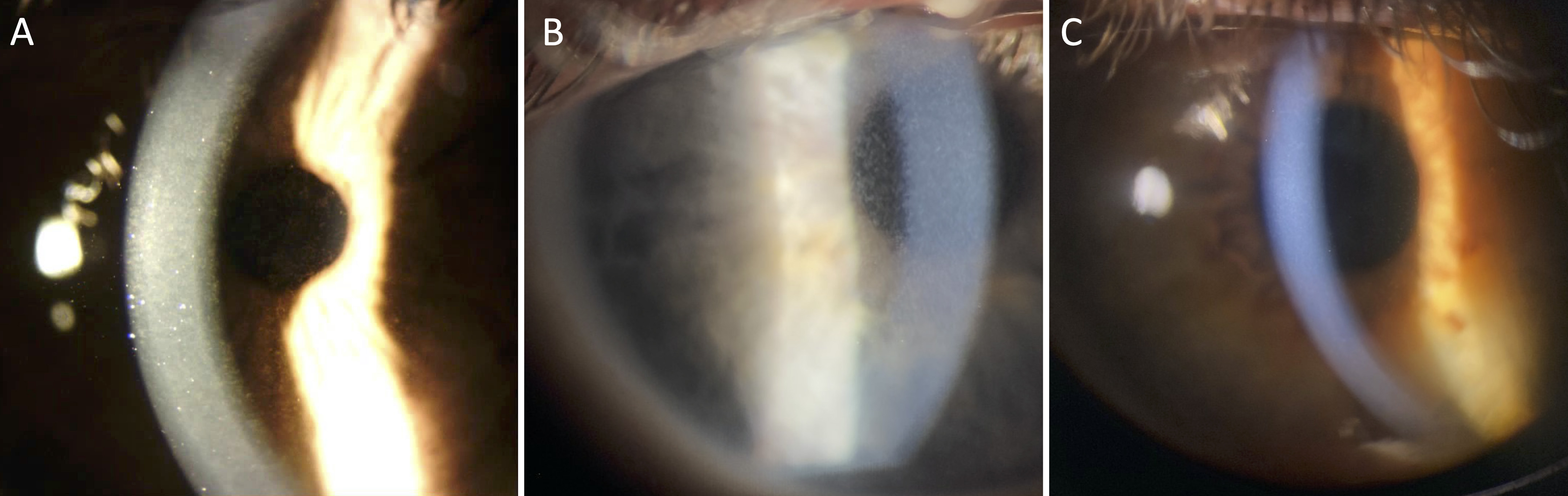
 Figure 1 of
Williams, Mol Vis 2023; 29:25-30.
Figure 1 of
Williams, Mol Vis 2023; 29:25-30.