Figure 3. ERK1/2 signaling promoted LEC proliferation in response to moderate H2O2 stimulus. A: Western blotting of p-ERK1/2, t-ERK1/2, and PCNA. B: EdU staining of the lens epithelial cells (LECs). ERK1/2 signaling was inactivated with pretreatment of LECs with 20 μM
of U0126 for 2 h before exposure to 50 μM of H2O2 for 12 h. Statistical analysis was performed with the paired Student t test (*p<0.05 and **p<0.01). The data are shown as mean ± standard error of the mean (SEM) with n = 3.
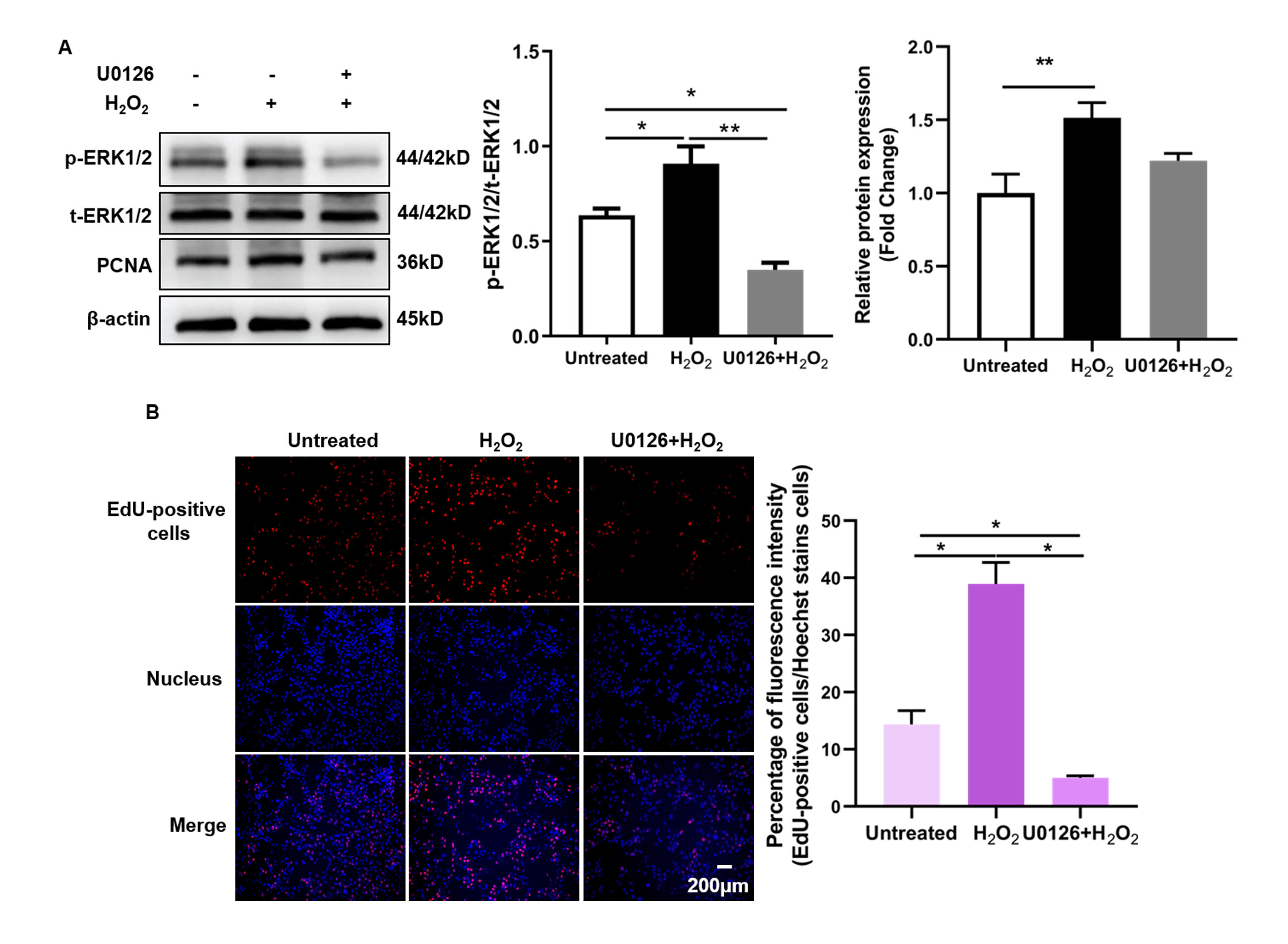
 Figure 3 of
Guo, Mol Vis 2023; 29:206-216.
Figure 3 of
Guo, Mol Vis 2023; 29:206-216.