Figure 2. Exposing LECs to 50 μM of H2O2 for 48 h activated Wnt/β-catenin signaling and promoted EMT. A: Western blotting of E-cadherin, vimentin, α-SMA, and Snail. B: Western blotting of Wnt 10a, p-LRP6Ser1490, p-GSK-3βser9, and β-catenin. C: Immunofluorescent images of β-catenin. Statistical analysis was performed with the paired Student t test (*p<0.05 and **p<0.01). The data are shown as mean ± standard error of the mean (SEM) with n = 3.
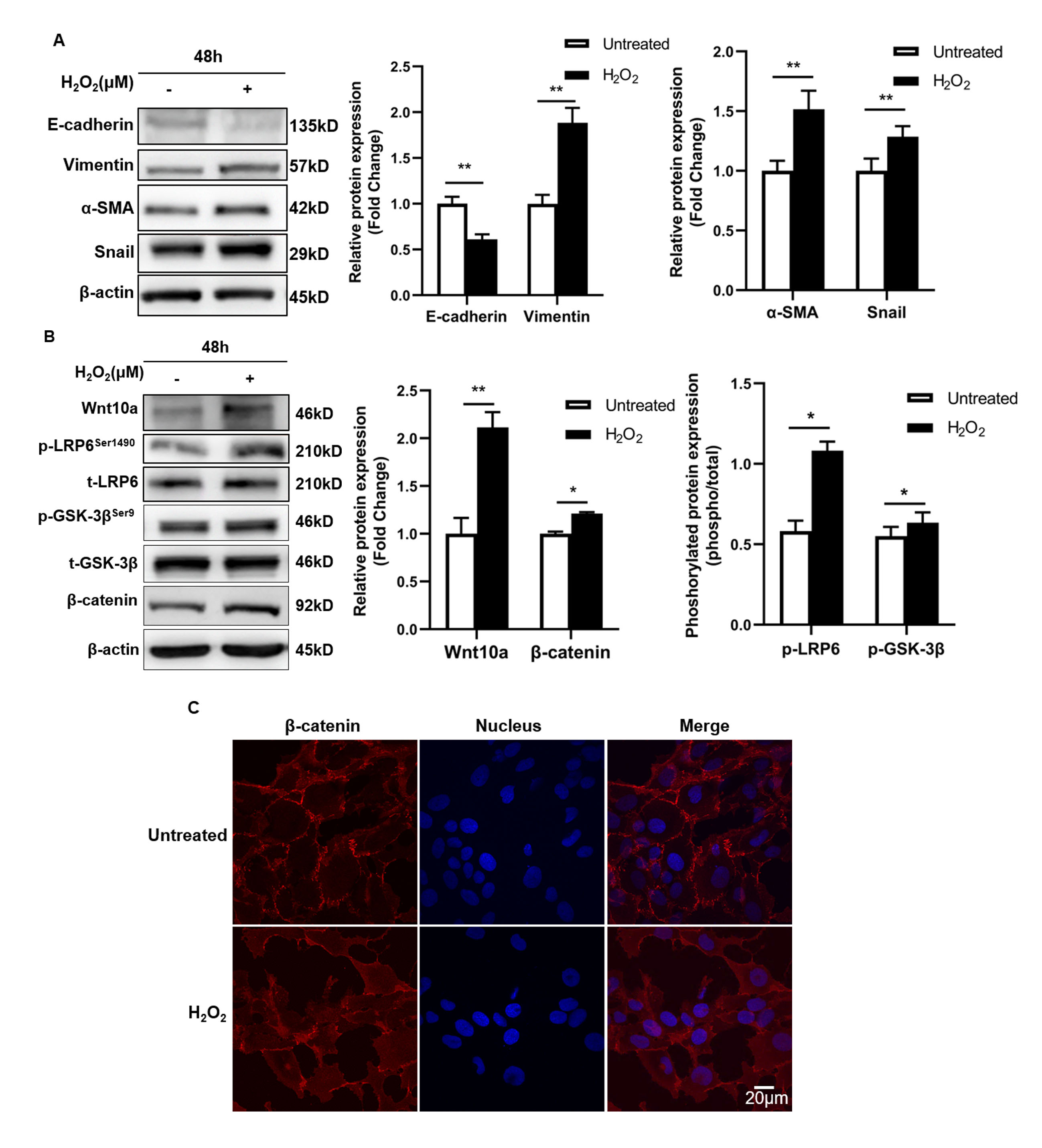
 Figure 2 of
Guo, Mol Vis 2023; 29:206-216.
Figure 2 of
Guo, Mol Vis 2023; 29:206-216.