Figure 1. Exposing LECs to moderate H2O2 resulted in the upregulation of phosphorylated ERK1/2 at both early and late time points. A: Western blotting of p-ERK1/2 and t-ERK/2 in lens epithelial cells (LECs) after treatment with 50 μM of H2O2 for 5, 10, 15, 30, and 60 min. Statistical analysis of the p-ERK1/2 to t-ERK1/2 ratio at different time points was performed
with one-way ANOVA followed by Dunnett’s multiple comparisons test (*p<0.05, **p<0.01, and ***p<0.001). B: Immunofluorescent staining of p-ERK1/2 after exposing LECs to 50 μM of H2O2 for 10 min. C: Western blotting of p-ERK1/2 and t-ERK1/2 after treatment of LECs with 50 μM of H2O2 for 1, 2, 4, 8, 12, 24, and 48 h. Statistical analysis of the p-ERK1/2 to t-ERK1/2 ratio was performed with the Student t test. The data are shown as mean ± standard error of the mean (SEM) with n = 3.
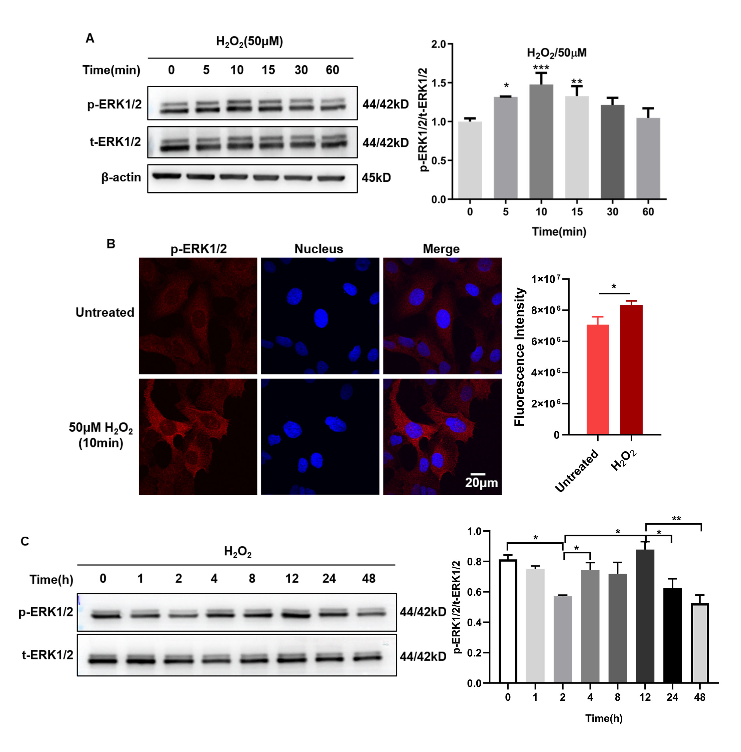
 Figure 1 of
Guo, Mol Vis 2023; 29:206-216.
Figure 1 of
Guo, Mol Vis 2023; 29:206-216.