Figure 4. Predicted protein effect of LTBP2 variants on structure–function properties. A–C (first column) show the wild-type in p.C207,
p.C1151, and p.C1372. A–C (second column) correspond to the variants of the wild-type described above. The disulfide bond
is indicated by the yellow line. The aromatic nucleus is blue. D indicates the predicted overall protein structure of wild-type,
p.W417X, and p.W1587X in brown. The transparent part indicates large fragment truncation.
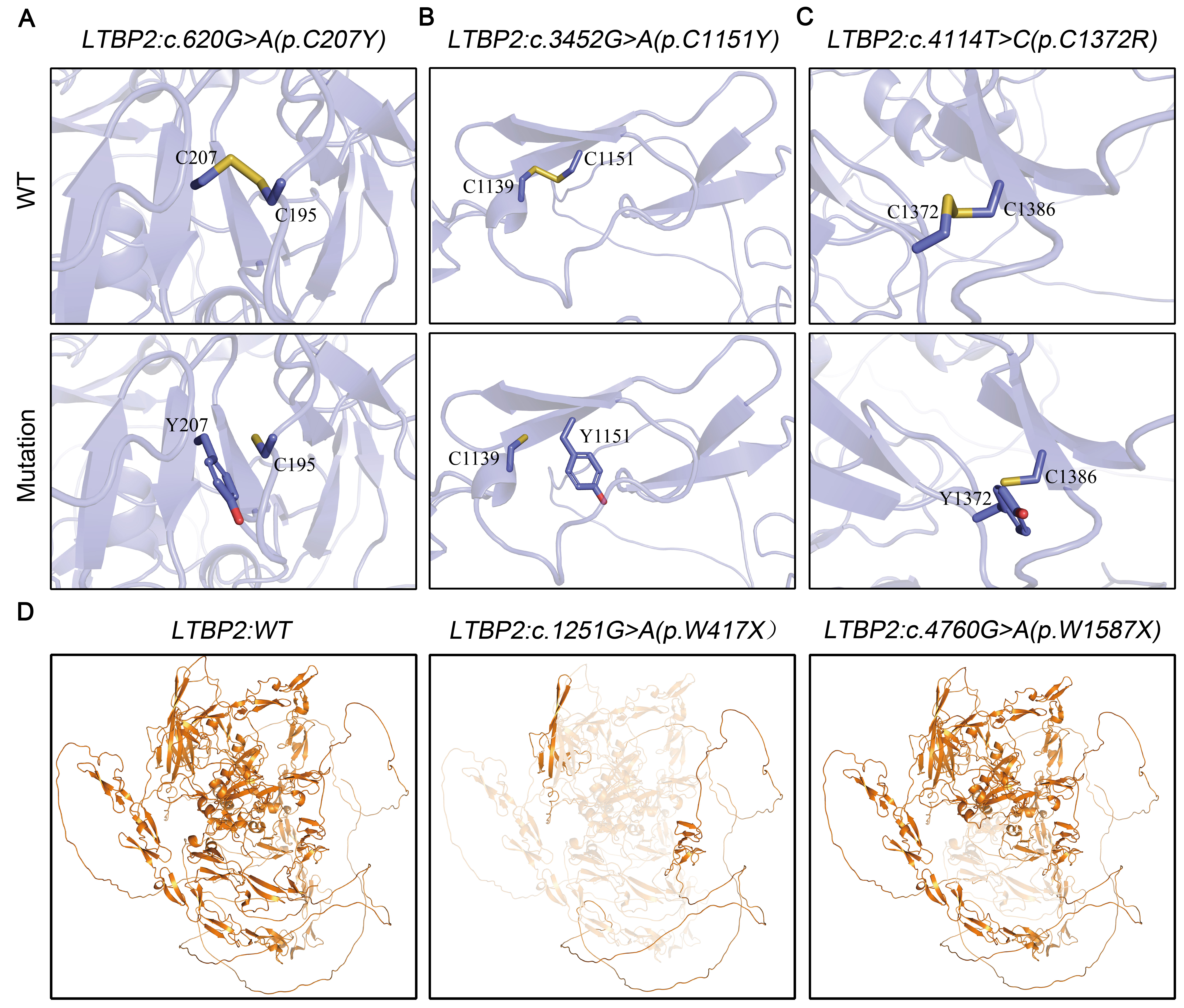
 Figure 4 of
Liu, Mol Vis 2023; 29:169-179.
Figure 4 of
Liu, Mol Vis 2023; 29:169-179.