Figure 2. Representative ocular clinical manifestations and clinical examinations of patients with LTBP2 mutations. A: The eye appearance
of Patient ID431 reveals a megalocornea feature. B: The slit-lamp photography of Patient ID431 illustrating an ectopia lentis
(EL). C: EL, coloboma lentis, and persistent pupillary membrane of Patient ID382. D: Anterior segment photography of Patient
ID294 depicts spherophakia and segmental iris atrophy (right eye). E: A fundus examination of patient ID382 shows a high cup/disc
ratio; 0.95 in the left eye. F: Fundus photography of Patient ID382 hints of optic disk abnormality.
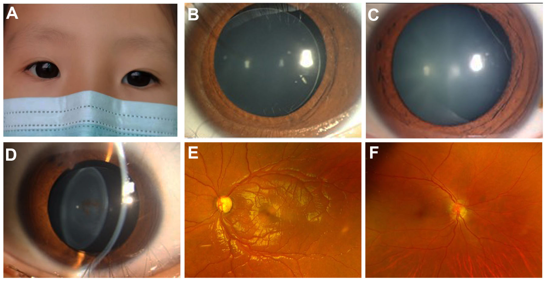
 Figure 2 of
Liu, Mol Vis 2023; 29:169-179.
Figure 2 of
Liu, Mol Vis 2023; 29:169-179.