Figure 1. Gene and protein structure of Basigin gene products expressed in the neural retina. A: An illustration of the gene structure is shown. Exons are represented by colored rectangles, and introns are represented
by the gray line. Exons are numbered and color-coded to show sequences translated into the extracellular immunoglobulin (Ig)
domains. B: An illustration of the protein structures of basigin-1 and basigin-2. Although they are depicted on the same membrane, they
are expressed by different cell types in the neural retina. Exon 1 (black) contains the leader sequence found in both proteins.
Exon 2, shown in purple, codes for the Ig0 domain specific to basigin-1. Exon 3, shown in green, codes for the Ig1 domain
found in both proteins. Exon 4, shown in green and orange, codes for Ig1 and Ig2, found in both proteins. Exon 5, shown in
orange, codes for Ig2. Exon 6 contains the coding sequence for the transmembrane domain. Exons 6, 7, and 8 (all black) code
for amino acids contained in both proteins and correspond to regions proximal to the membrane, the transmembrane domain, and
the cytoplasmic domain, respectively.
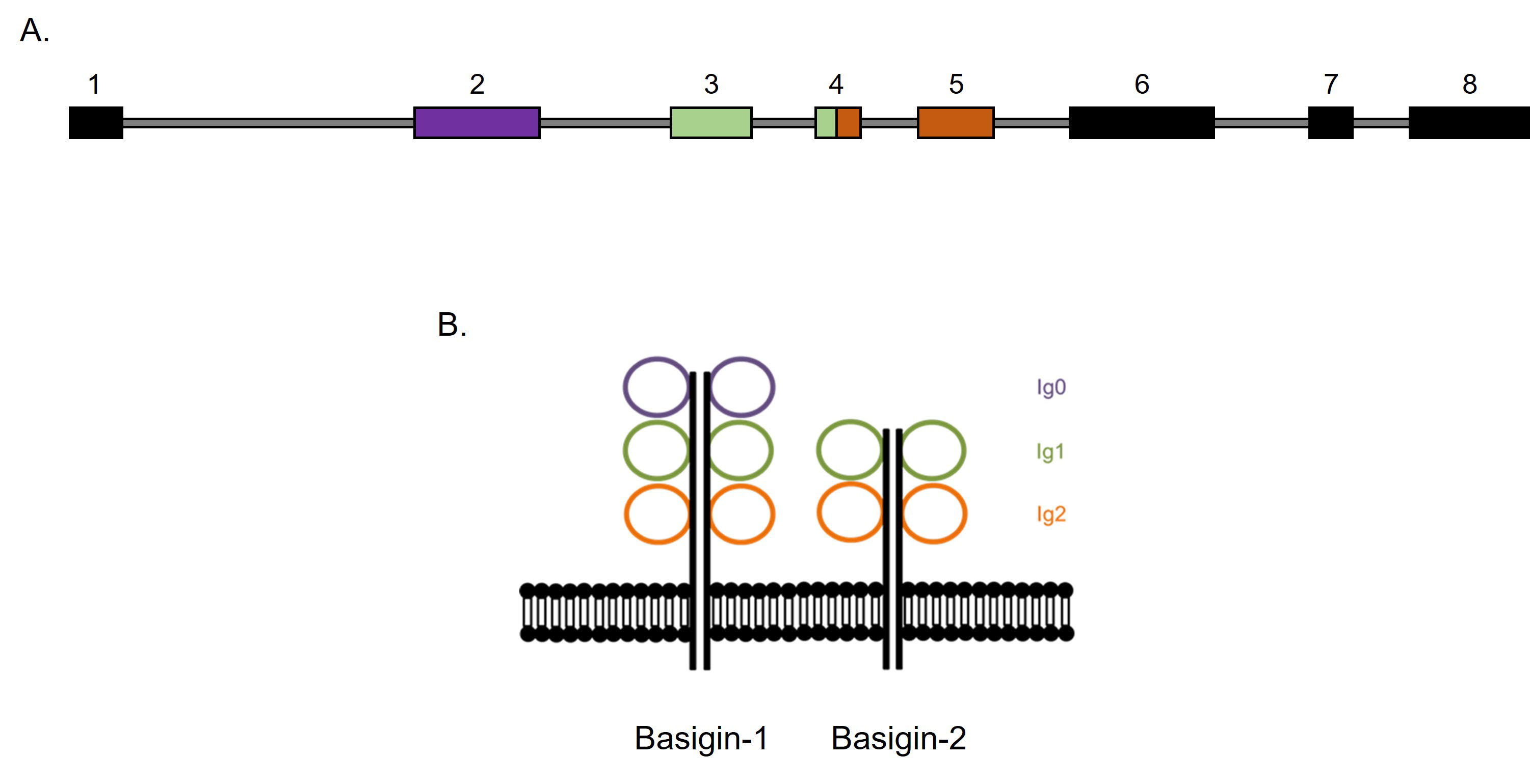
 Figure 1 of
Solstad, Mol Vis 2023; 29:13-24.
Figure 1 of
Solstad, Mol Vis 2023; 29:13-24.