 Appendix 4 of
Ben Yosef, Mol Vis 2023; 29:1-12.
Appendix 4 of
Ben Yosef, Mol Vis 2023; 29:1-12. Appendix 4 of
Ben Yosef, Mol Vis 2023; 29:1-12.
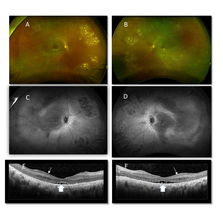
Appendix 4. Multimodal retinal imaging from patient 17 with ARB.
To access the data, click or select the words “Appendix 4.” A,B: Optos widefield fundus photos of right and left eye respectively, showing multiple yellow-white deposits in the macula and extramacular area and patches of retinal pigment epithelium (RPE) atrophy in the periphery, C, D: Widefield fundus autofluorescence photos, showing autofluorescence changes at the macula and mid periphery and temporal peripheral hypo-autofluorescent patches corresponding to the RPE atrophic patches. E, F: Spectral-domain OCT images showing cystoid macular intraretinal fluid (narrow arrow) and subretinal fluid with hyperreflective deposits (wide arrow).