Figure 2. Anterior eye photos of affected members in this study. Images of the anterior segment of both eyes of patients IV:1 (aged
48 years), IV:2 (aged 46 years), and IV:3 (aged 42 years) from family MEP68 (A), and the proband (aged 1 year) from family
F1332 (B). Note microcornea (A, IV:1 & IV:3), peripheral corneal opacity (A, IV:1 & IV:3), sclerocornea totalis (A, IV:2 & B), and iris coloboma (A, IV:1, right eye, arrowed).
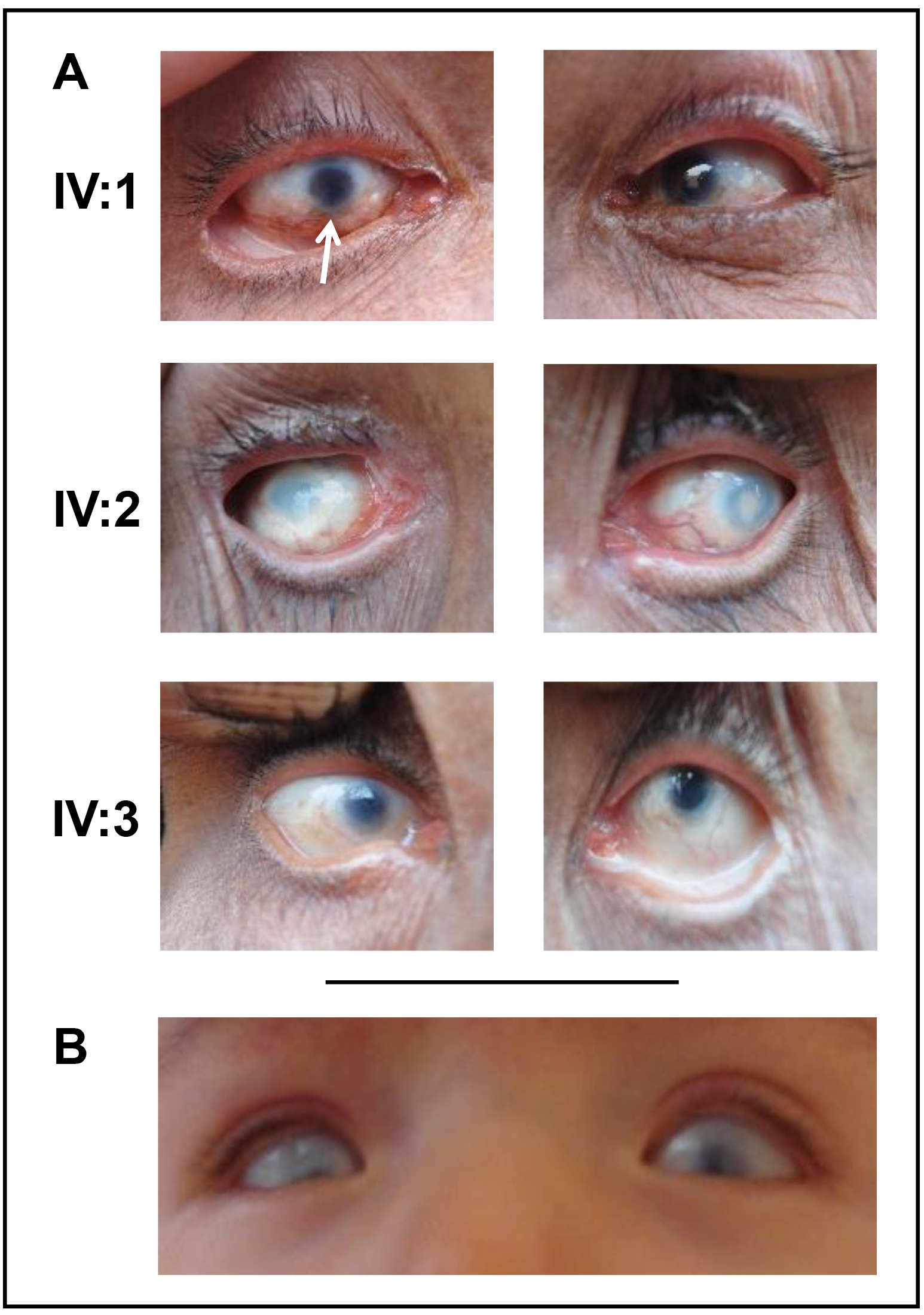
 Figure 2 of
Panagiotou, Mol Vis 2022; 28:57-69.
Figure 2 of
Panagiotou, Mol Vis 2022; 28:57-69.