Figure 7. SPS inhibited HSV-1 infection by blocking viral absorption. HCE-T cells were incubated with HSV-1g for 2 h at 4°C, and different
concentrations of SPS were then added during absorption (A) and penetration (B). GFP fluorescence during absorption (C) and
penetration (D) was detected using an inverted fluorescence microscope. The average fluorescence intensity was measured using
ImageJ software. The average intensity of the control cells was assigned a value of 100. Data are presented as means ± standard
deviations (n = 3). **p < .01 compared with the control group. *p < .05 compared with the control group.
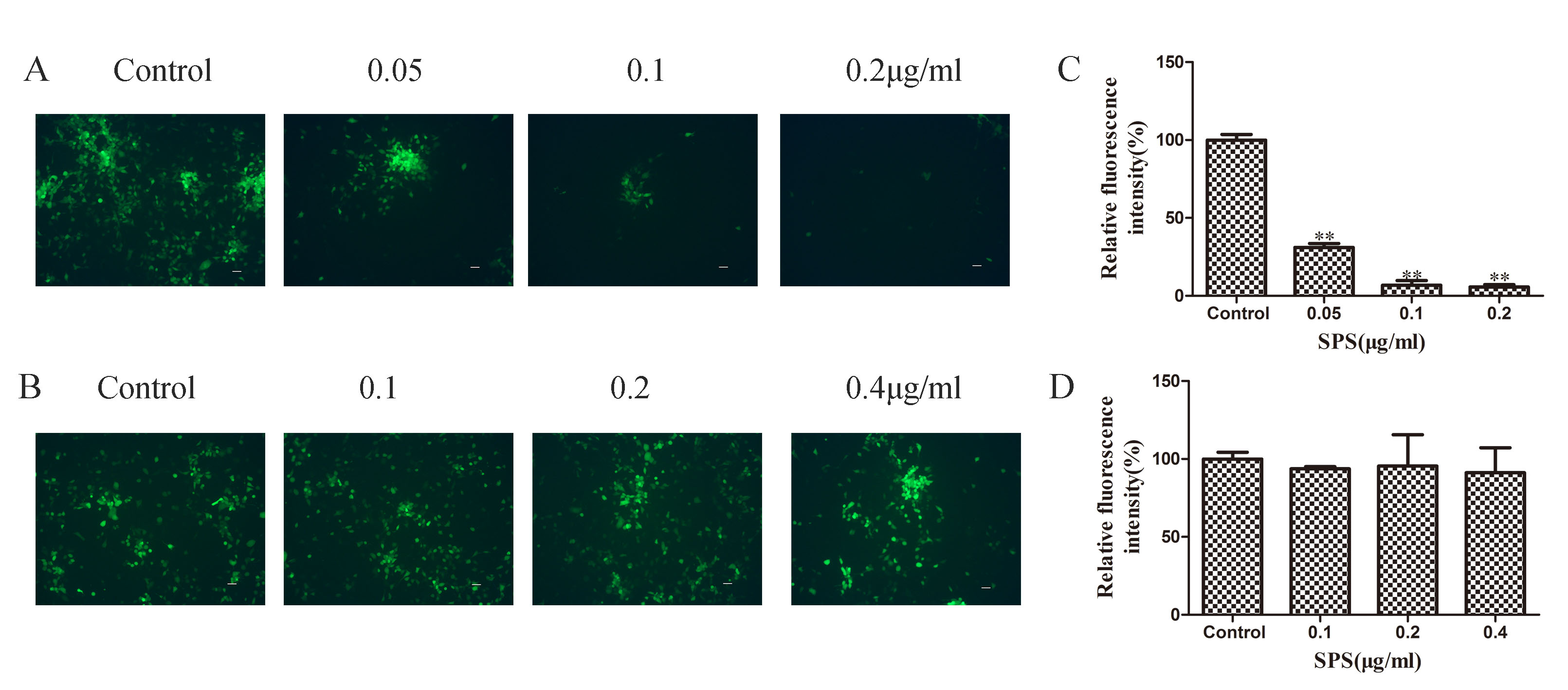
 Figure 7 of
Li, Mol Vis 2022; 28:516-525.
Figure 7 of
Li, Mol Vis 2022; 28:516-525.