Figure 2. The antiviral effect of SPS on preincubated HCE-T cells. HCE-T cells were preincubated with various concentrations of SPS
for 1 h at 37°C and then subjected to washing (A) or no washing (B) with PBS before inoculation with HSV-1g. After 24 h, GFP
fluorescence was detected using fluorescence microscopy and quantified using ImageJ software (C). The average intensity of
the control cells was assigned a value of 100. Values are presented as means ± SD (n = 3). **p < .01 compared with the control group.
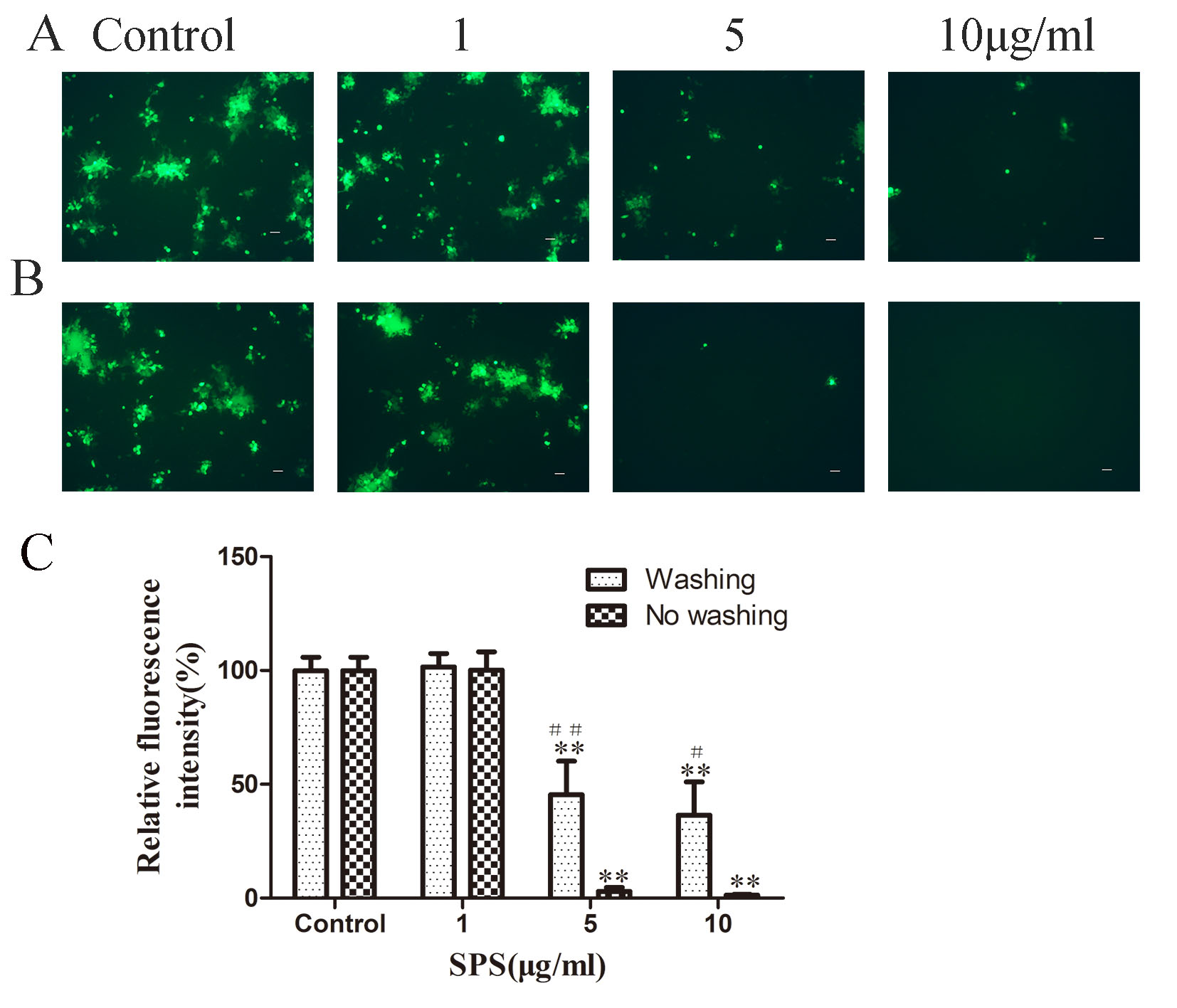
 Figure 2 of
Li, Mol Vis 2022; 28:516-525.
Figure 2 of
Li, Mol Vis 2022; 28:516-525.