Figure 5. The expression of TSPO in normal retina and blue light–damaged retina. A: The expression and location of translocator protein (TSPO) were analyzed with immunohistochemical staining. B: Immunofluorescence staining of the retinal slices. (TSPO: red fluorescence, IBA-1 (a microglia marker): green fluorescence,
2-(4-Amidinophenyl)-6-indolecarbamidine dihydrochloride (DAPI): blue fluorescence). C: The expression of TSPO in the retina was analyzed with western blotting. **p<0.01 versus the control group. INL: inner nuclear
layer, ONL: outer nuclear layer, GCL: ganglion cell layer.
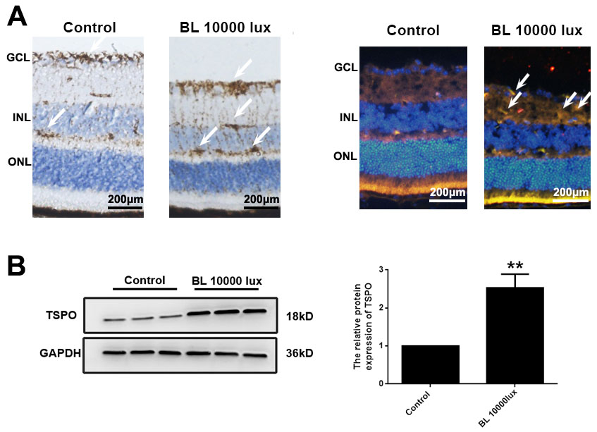
 Figure 5 of
Chen, Mol Vis 2022; 28:507-515.
Figure 5 of
Chen, Mol Vis 2022; 28:507-515.