Figure 1. Clinical features of patient EVR-20 with Norrie disease. A: Summary of ocular findings. B: Anterior segment of the right eye with cataract, corneal clouding, and posterior synechiae. C: Anterior segment of the left eye with cataract and posterior synechiae. D: Ocular ultrasound of the right eye shows the density of vitreous opacities distributed almost throughout the vitreous body.
E: Ocular ultrasound of the left eye shows a thick membrane attached to the optic disc and the funnel configuration of retinal
detachment. OD (oculus dexter): right eye; OS (oculus sinister): left eye; mm: millimeters; mm Hg: millimeters of mercury; +: yes; -: no.
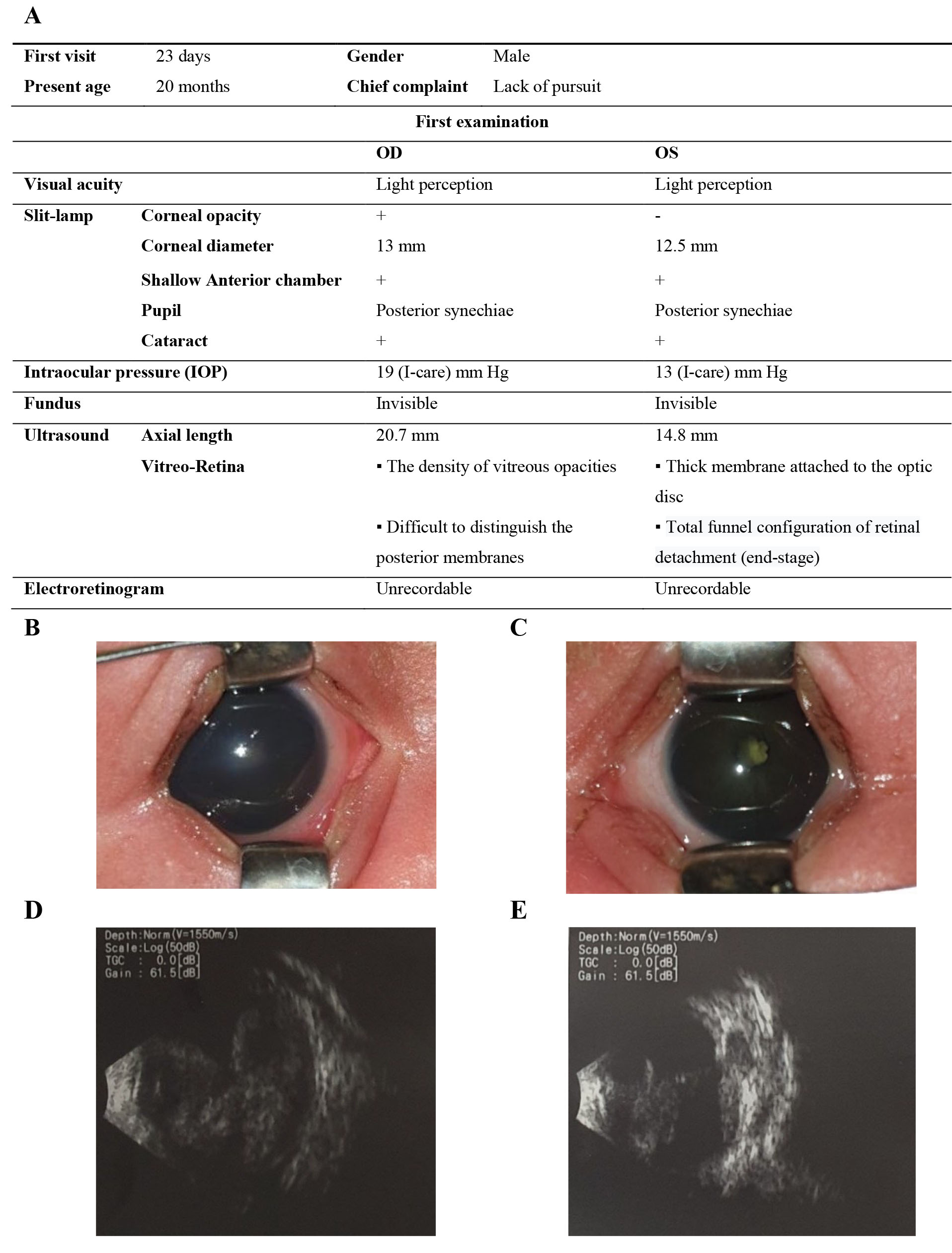
 Figure 1 of
Trang, Mol Vis 2022; 28:480-491.
Figure 1 of
Trang, Mol Vis 2022; 28:480-491.