Figure 8. Photographs of the eye anterior segment in rats in the NT group (A, C) and Mino group (B, D) at one and three months after beginning the eye drop administration. A and B show cataract development in diabetic rats
at one month after beginning the eye drop administration. C and D show iris vessel proliferation, thickening, and tortuosity (white arrows) three months after beginning the eye drop administration.
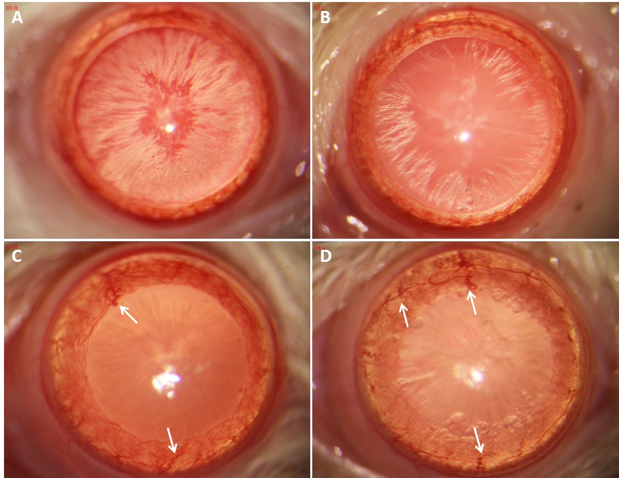
 Figure 8 of
Li, Mol Vis 2022; 28:460-479.
Figure 8 of
Li, Mol Vis 2022; 28:460-479.