Figure 4. RGCs and cells of the outer nuclear layer are shown with electron microscopy. A, D: The retinal ganglion cell (RGCs) and the cells of the outer nuclear layer have a defined plasma membrane and uniformly distributed
chromatin in the control group. B, E: There are many autophagosomes (black arrow) in the oxygen-induced retinopathy (OIR) group. C, F: There are fewer autophagosomes (black arrow) in the human adipose mesenchymal stem cell (hADSC) injection group than in
the OIR and control groups. However, there is no noticeable difference in cells among the three groups.
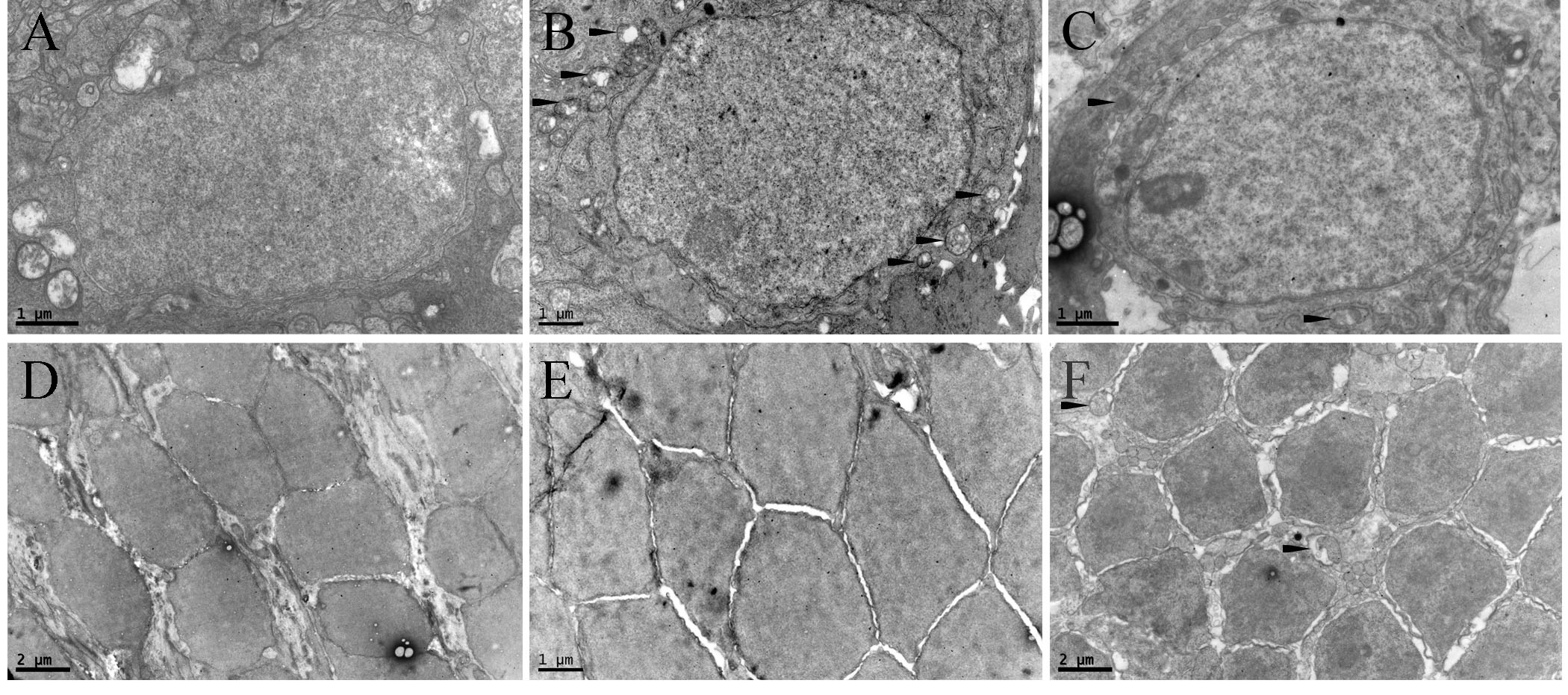
 Figure 4 of
Zhou, Mol Vis 2022; 28:432-440.
Figure 4 of
Zhou, Mol Vis 2022; 28:432-440.