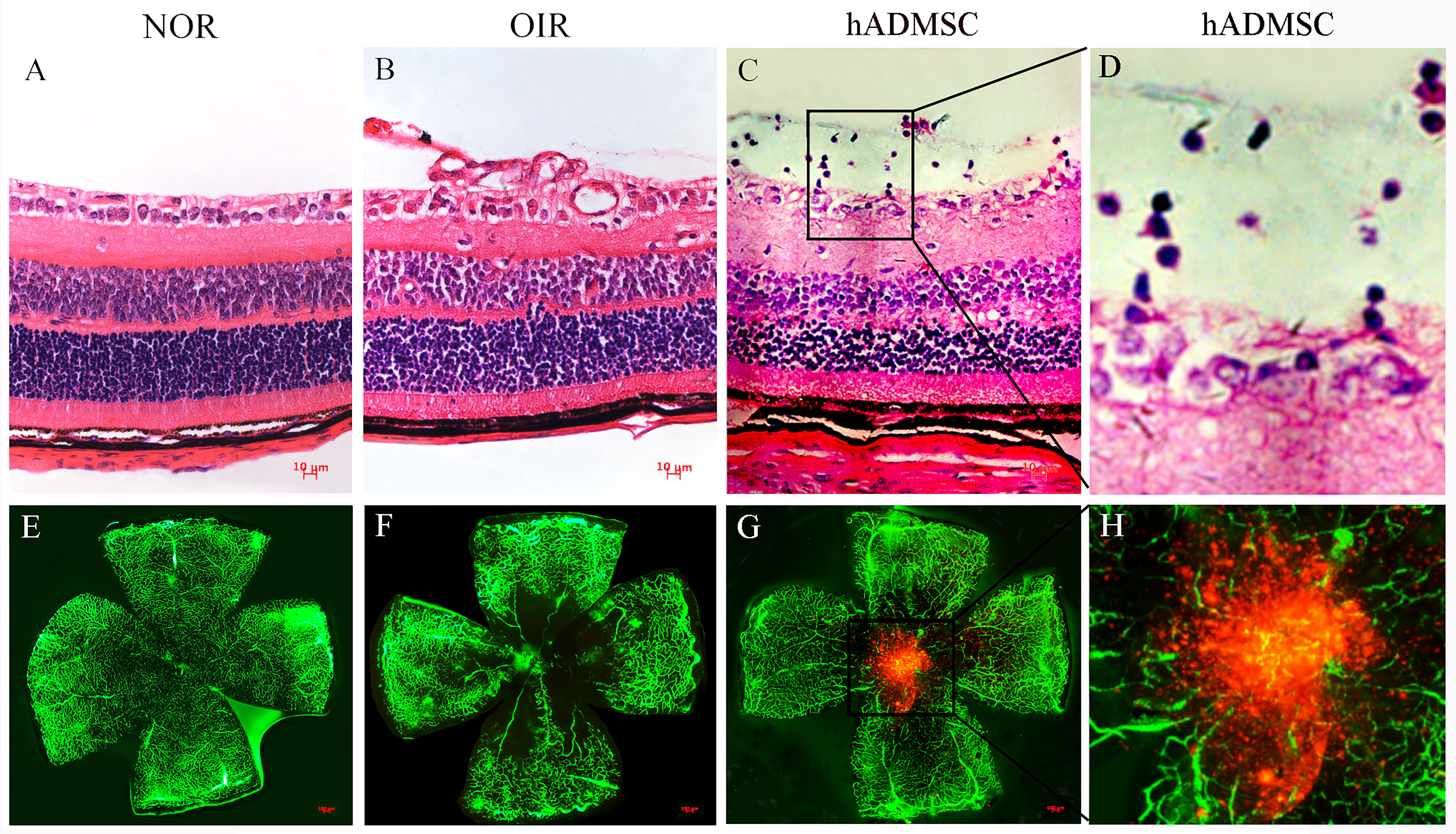
Figure 2. Intraocular tracking of hADSCs.
A: The structure of each layer of the retina is clear, and there is no neovascularization breaking through the internal limiting
membrane (ILM) in the P17 control group.
B: Extensive neovascularization broke through the ILM in the P17 oxygen-induced retinopathy (OIR) group.
C: No apparent neovascularization breaking through the ILM and the presence of injected cells above the ILM (not fused with
the retina) are noted in the human adipose mesenchymal stem cells (hADSCs) injection group.
D: Partial magnification of
Figure 3C (30X).
E: The retinal blood vessels are smooth and without neovascularization, and there is no perfusion area in the control group.
F: There is extensive highly green fluorescent neovascularization in the periphery area and non-perfusion in the central area
of the retina in the OIR group.
G: Neovascularization and non-perfusion are significantly reduced in the hADSC injection group compared with that in the OIR
group, and labeled hADSCs with red fluorescence are seen above the neovascularization and non-perfusion area.
H: Partial magnification of
Figure 3G (30X).
 Figure 2 of
Zhou, Mol Vis 2022; 28:432-440.
  Figure 2 of
Zhou, Mol Vis 2022; 28:432-440.