Figure 1. Culture, labeling, and immunofluorescence staining of hADSCs. A: In the second passage, the human adipose mesenchymal stem cells (hADSCs) show spindle and polygonal shapes. B: The membrane of the hADSCs shows red fluorescence, and the nucleus shows no fluorescence. C: After induction, the corners of the hADSCs appear rounded. D, E, F: The expression of von Willebrand Factor (vWF) is absent (no fluorescence) in the control group. G, H, I: The expression of vWF is present (green fluorescence) in the experimental group. The nuclei stained with 4′,6-diamidino-2-phenylindole
(DAPI) shows blue fluorescence in each group. Scale bar = 20 µm, 200X; scale bar = 10 µm, 400X.
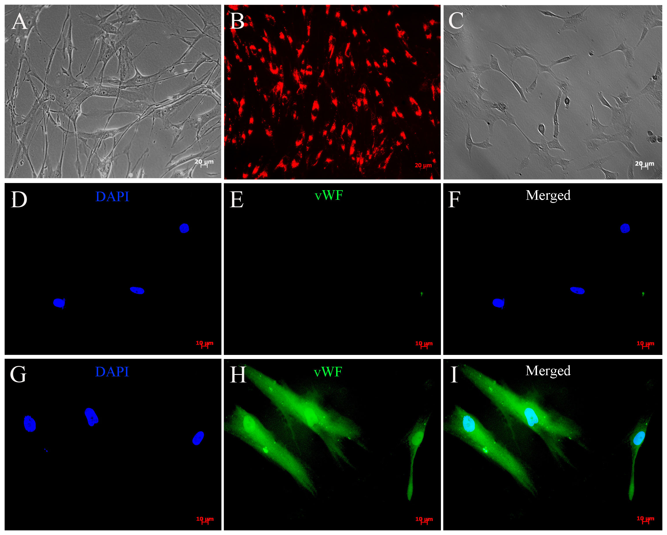
 Figure 1 of
Zhou, Mol Vis 2022; 28:432-440.
Figure 1 of
Zhou, Mol Vis 2022; 28:432-440.