Figure 3 of
Shamsnajafabadi, Mol Vis 2022; 28:412-431.
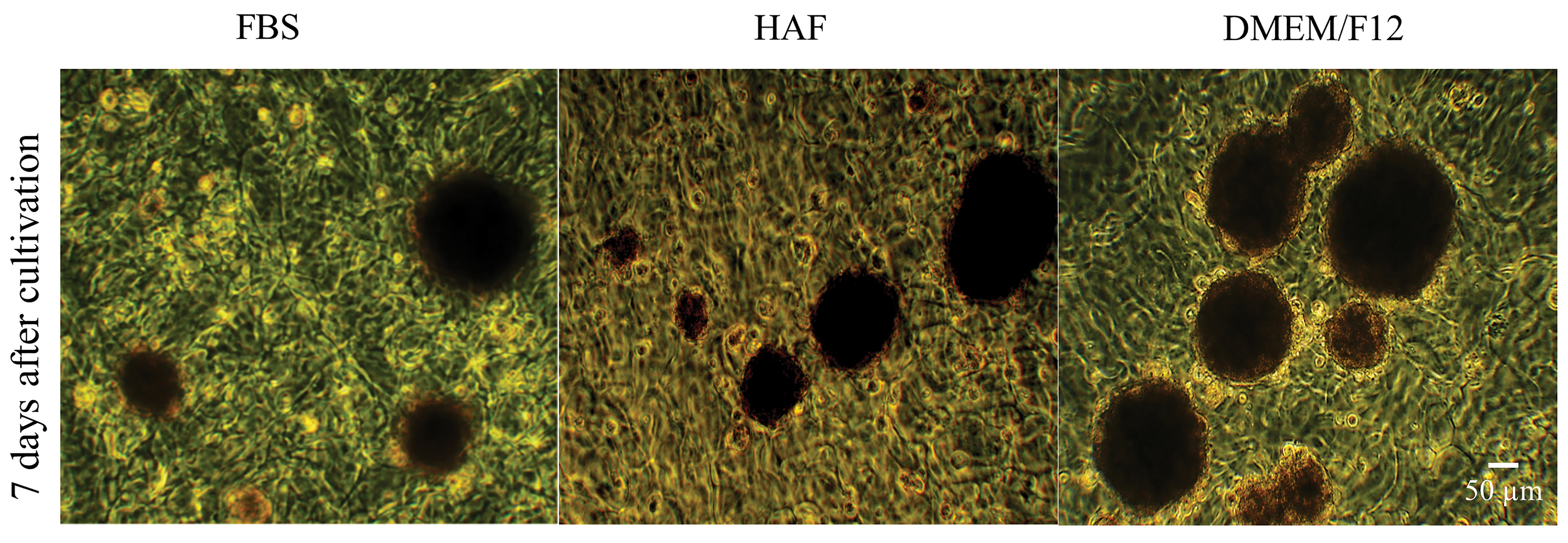
Figure 3.
Phase-contrast micrograph of hRPE cells (Passage 4), on A/G substrate. The dense spheroids developed on A/G substrate in FBS, HAF and DMEM/F12 -treated cultures 7 days after cultivation. Magnification: 200X.
 Figure 3 of
Shamsnajafabadi, Mol Vis 2022; 28:412-431.
Figure 3 of
Shamsnajafabadi, Mol Vis 2022; 28:412-431.