Figure 2. Phase-contrast micrograph of hRPE cell morphology on polystyrene (Passage 4) under the treatment of FBS, DMEM/F12 and HAF.
Elongated fusiform morphology of the cells developed in FBS- and DMEM-treated cultures. The aforesaid structures formed in
HAF-treated cultures on the first day of starting the cultures, whereas the floated and attached spheroids appeared on the
7th day of the cultures. Magnification: 200X.
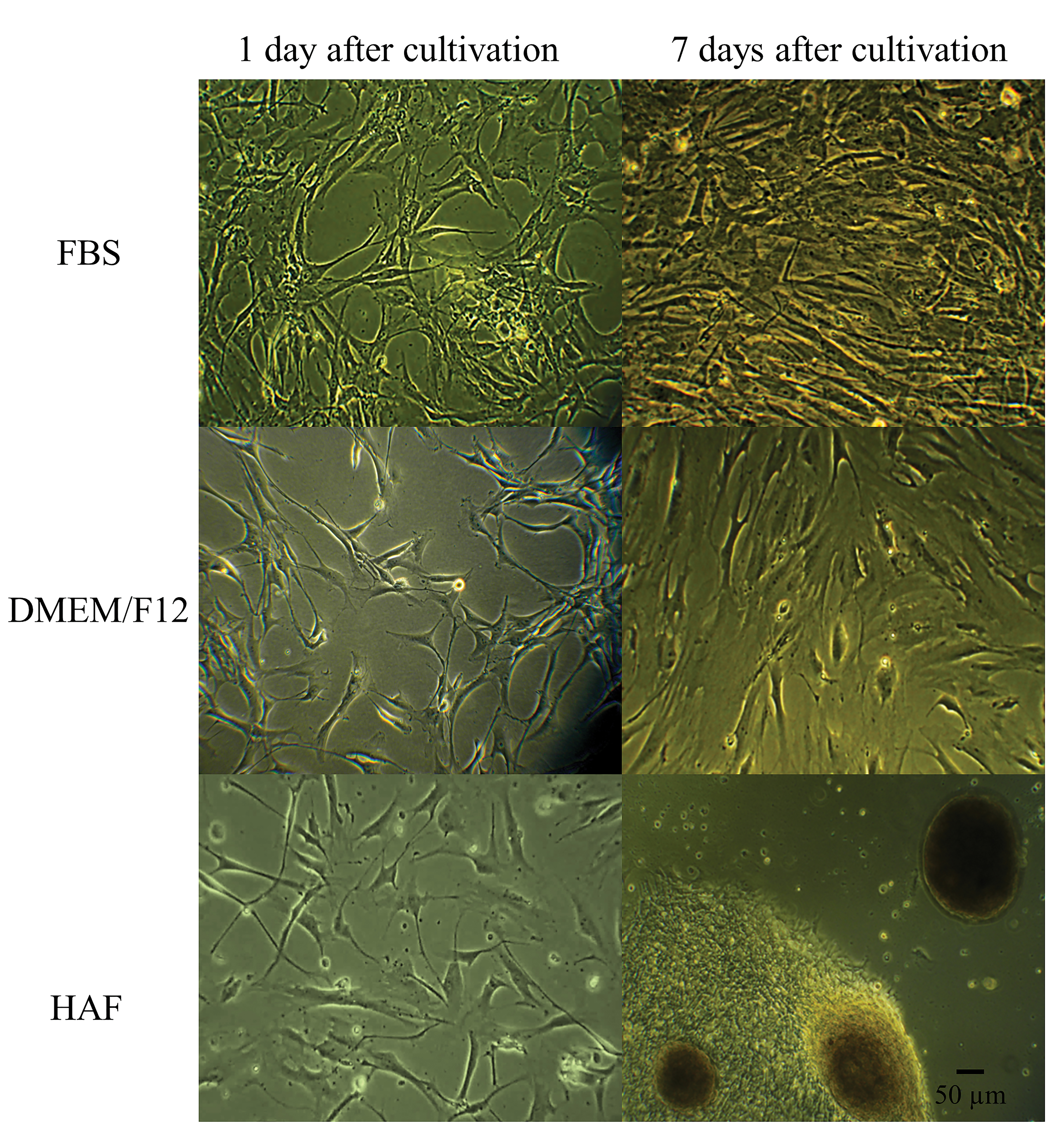
 Figure 2 of
Shamsnajafabadi, Mol Vis 2022; 28:412-431.
Figure 2 of
Shamsnajafabadi, Mol Vis 2022; 28:412-431.