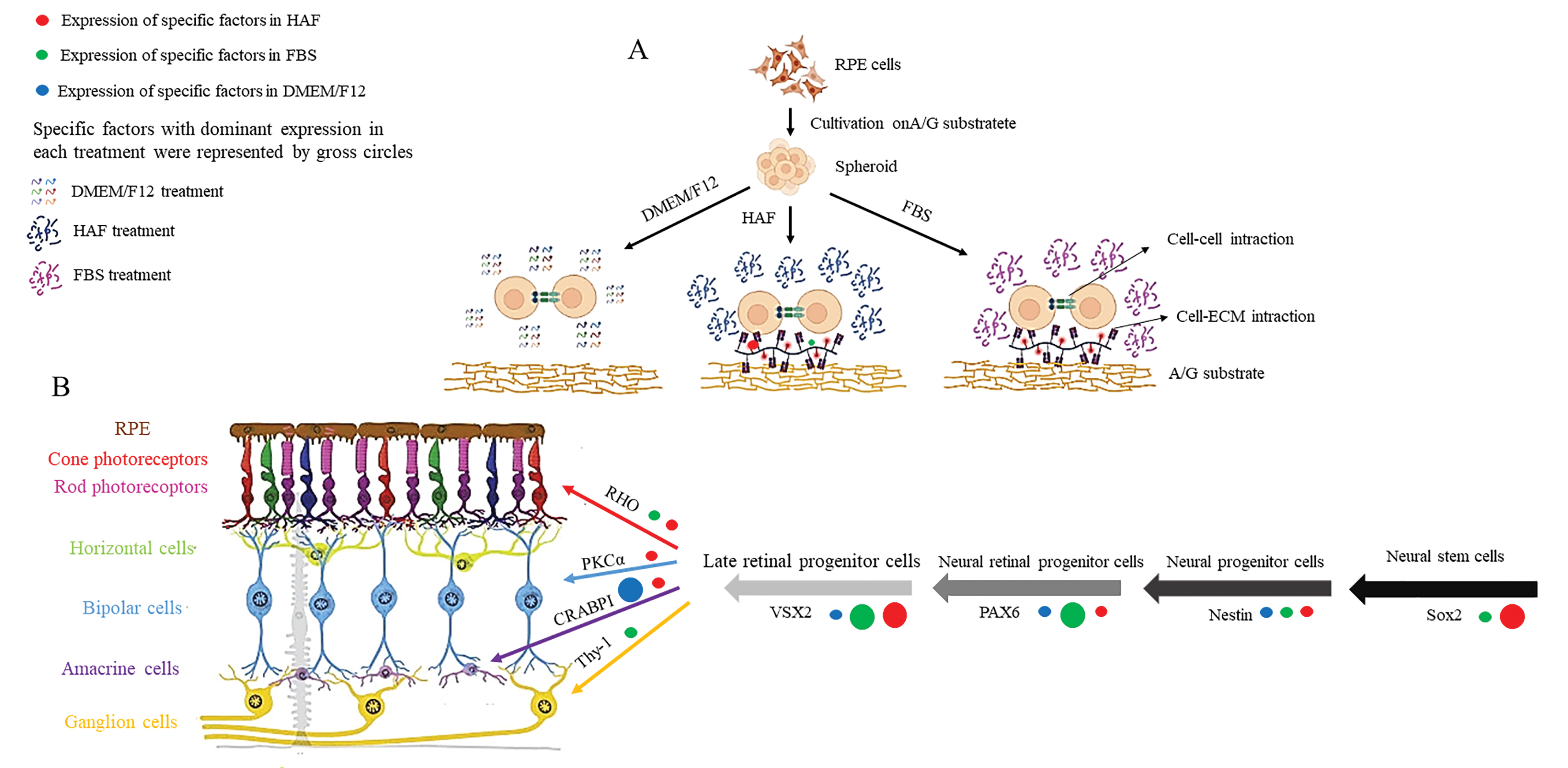
Figure 10. hRPE cells on A/G substrate.
A: Schematic presentation of hRPE cell cultivation on A/G substrate. hRPE cells formed spheroids when they were cultured on
A/G substrate. A/G substrate facilitated cell–ECM interactions when cultures were supplemented with HAF or FBS. In a DMEM/F12-treated
cultures, A/G substrate directed hRPE cells to establish cell–cell interactions.
B: Schematic presentation of stem cell differentiation into neural retinal cell lineage. The expression patterns of specific
markers during differentiation are presented. Factors that are expressed in mRPE cell cultures under treatment with HAF, FBS,
or DMEM/F12 are represented by red, green, and blue circles, respectively. Specific factors with dominant expression in each
treatment are represented by gross circles. Figure was created with
BioRender.
 Figure 10 of
Shamsnajafabadi, Mol Vis 2022; 28:412-431.
Figure 10 of
Shamsnajafabadi, Mol Vis 2022; 28:412-431.