Figure 1. Phase-contrast micrograph of hRPE cells’ primary culture morphologies, immunostaining for verification of the cells’ identity.
A: Polygonal cells with densely pigmented morphology of hRPE cells 1 day after cultivation. B: 3 days later, hRPE cells formed a monolayer of elongated fusiform cells. C: They made small, tightly packed cobblestone structures with distinct phase-bright borders in confluent cultures 7 days after
cultivation. D: Merged image for the RPE65 marker (green) and DAPI nuclear staining (blue). Immunocytochemistry analysis in the 5th passage, represented that, more that 90% of hRPE cells in the population were positive for RPE65. Magnification: (A, B, and
C: 320X and D: 400X).
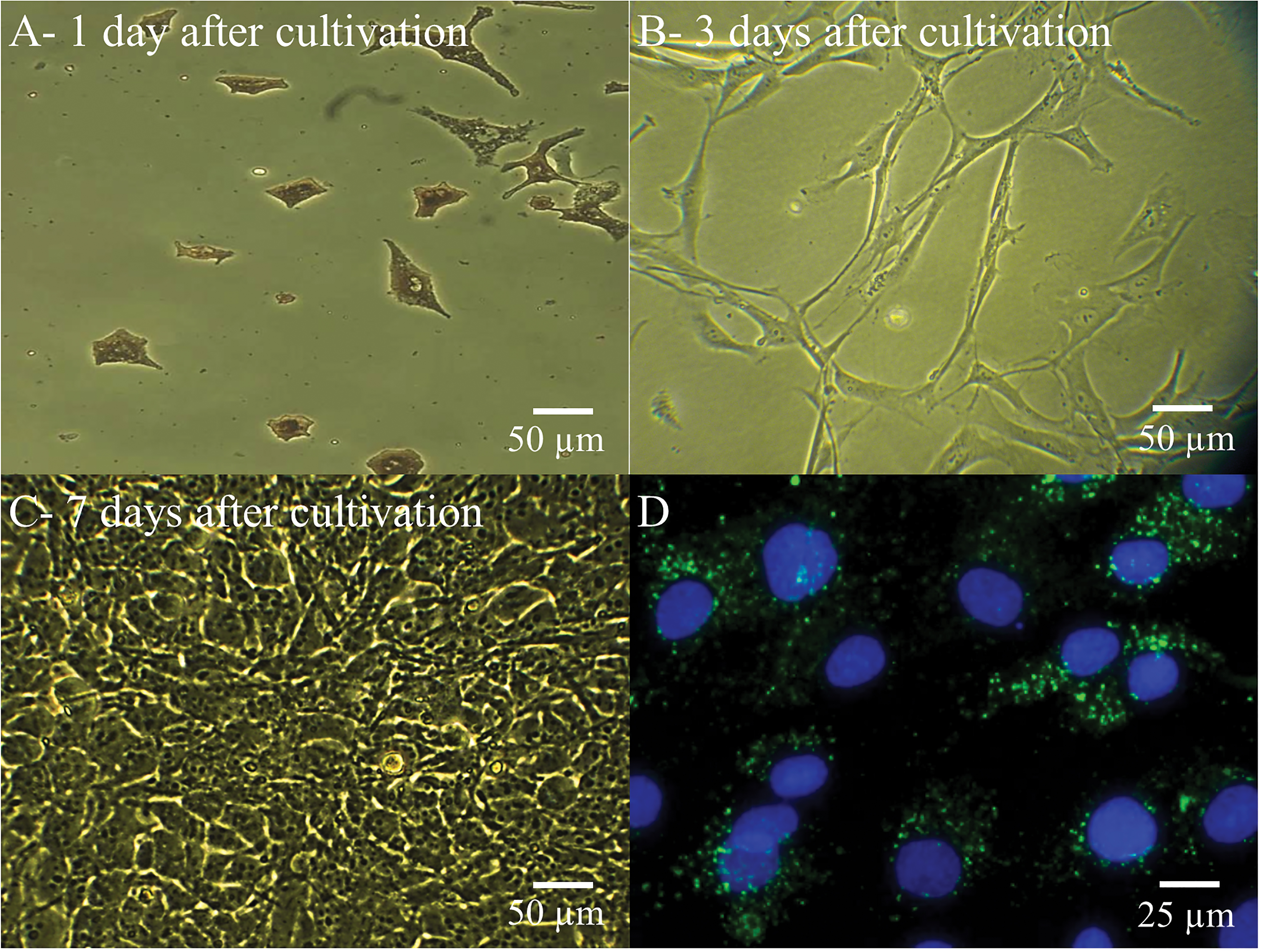
 Figure 1 of
Shamsnajafabadi, Mol Vis 2022; 28:412-431.
Figure 1 of
Shamsnajafabadi, Mol Vis 2022; 28:412-431.