Figure 4. Effect of presence or absence of exogenous Chol in the culture medium of DYS, CWI, and A2 RPE cells on cellular sterol profiles.
7DHC/Chol mole ratios were quantified by reverse-phase HPLC analysis of non-saponifiable lipids extracted from cells cultured
for 9 days in medium containing either 0.9% (v/v) bovine calf serum (BCS) or delipidated (sterol-deficient) calf serum (DLCS).
Upper panels: 7DHC/Chol mole ratios (average values, n = 3/each condition/cell line) are expressed as a function of the culture condition.
A2 RPE cells exhibited the highest levels of 7DHC, enhanced by culturing in the DLCS-containing medium. When cultured in medium
containing BCS, the sterol profile of CWI RPE cells was comparable to that of DYS (control) RPE cells; however, 7DHC levels
increased significantly when CWI RPE cells were switched to DLCS-containing medium, whereas the DYS RPE cells did not respond
similarly (n = 3, *p <0.01, #p <0.05). Lower panels: Light microscopy images of Oil Red O-stained SLOS-derived (CWI and A2) RPE cells and control (DYS) RPE cells (n = 3/ cell
line). A2 RPE cells exhibited substantial accumulation of lipid droplets, while only select CWI RPE cells exhibited lipid
droplet accumulation (black arrows). By contrast, DYS RPE cells did not exhibit lipid droplet accumulation. Scale bar: 10 μm.
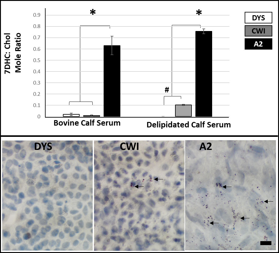
 Figure 4 of
Farkas, Mol Vis 2022; 28:394-411.
Figure 4 of
Farkas, Mol Vis 2022; 28:394-411.