Figure 4. SFN treatment can restore the retinal morphology disrupted by MnSOD knockdown. Postmortem histological sections (5
μ
m thick) of retinas prepared from WT and Nrf2 KO mice and labeled with hematoxylin and eosin (H&E) staining. The images in
the top row are from WT eyes, and the images in the bottom row are from Nrf2 KO eyes. Sections were prepared from animals:
(A) untreated, (B) treated with SFN in the absence of MnSOD knockdown, (C) administered AAV1-RzSOD2 to knock down MnSOD, and (D) administered AAV1-RzSOD2 to knock down MnSOD and treated with SFN. Red arrows in (B) mark patchy areas of atrophy (top panel) and subretinal vacuoles (bottom panel). In the bottom panel of (C), red arrows denote severe retinal/RPE thinning and subretinal infiltrates.
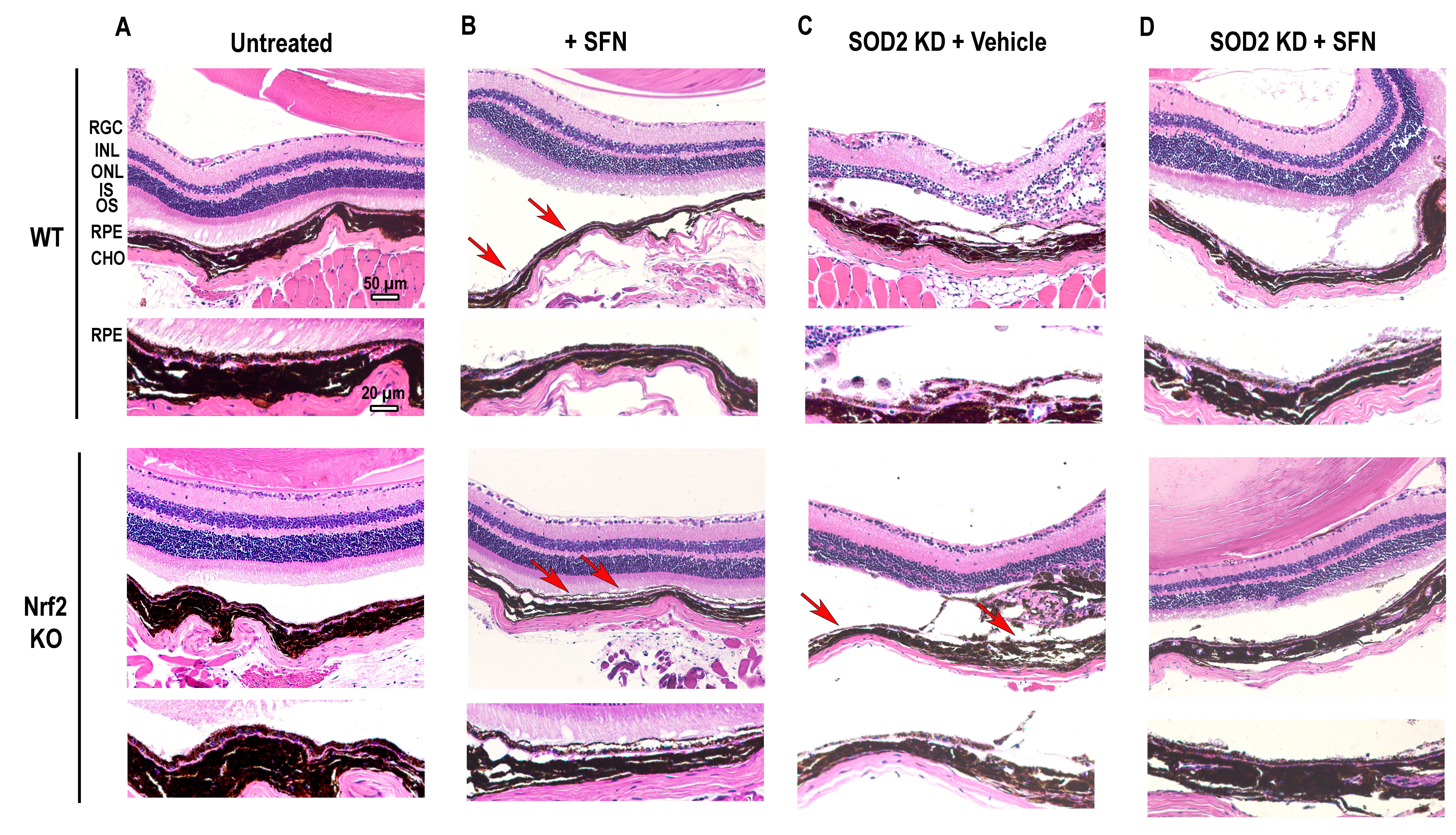
 Figure 4 of
Qi, Mol Vis 2022; 28:378-393.
Figure 4 of
Qi, Mol Vis 2022; 28:378-393.