Figure 4. Evaluation of the effect of PRPF31 c.1146+5G>T on splicing in vivo. A: RT-PCR analysis of a lymphocyte-derived RNA sample with primers in exons 10 and 12. The genotypes of the family members
are indicated above each lane. B: Evaluation of the relative abundance of alternatively spliced transcripts using amplicon-based next-generation sequencing.
Shown is the number and percentage of exon 11+ compared with those of exon 11− unique reads in each tested family member.
C: Average frequency of exon 11− transcripts in the wt and heterozygous (het) individuals. +, wt; M, mutant. *p < 0.05.
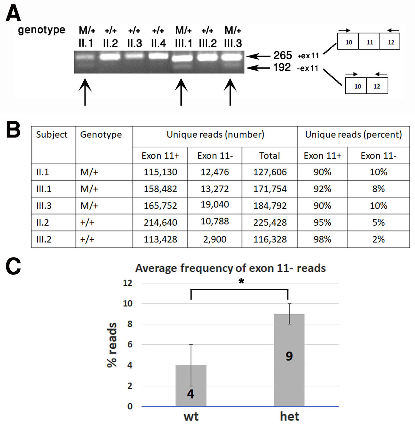
 Figure 4 of
Ali-Nasser, Mol Vis 2022; 28:359-368.
Figure 4 of
Ali-Nasser, Mol Vis 2022; 28:359-368.