Figure 2. Clinical images of subjects II:1 and III:1. A: Clinical image of Subject II:1, showing retinal manifestations of outer retinal degeneration. The infrared scanning laser
ophthalmoscopy (SLO-IR; a, c) and spectral-domain optical coherence tomography (SD-OCT; b, d) images of each eye show a perifoveal
ellipsoid zone and outer nuclear layer loss, with preservation of these layers in the foveal center. Focal vitreomacular traction
with distortion of the foveal contour can be observed in the right eye, whereas subtle cystic changes can be observed in the
inner nuclear layer temporal to the fovea in the left eye. B: Macular image of Subject III:1. The infrared SLO-IR (a, c) and SD-OCT images (b, d) show preservation of the perifoveal
ellipsoid zone and outer nuclear layer in the fovea, with perifoveal thinning of these structures in each eye. Calibration
bars: 200 µm.
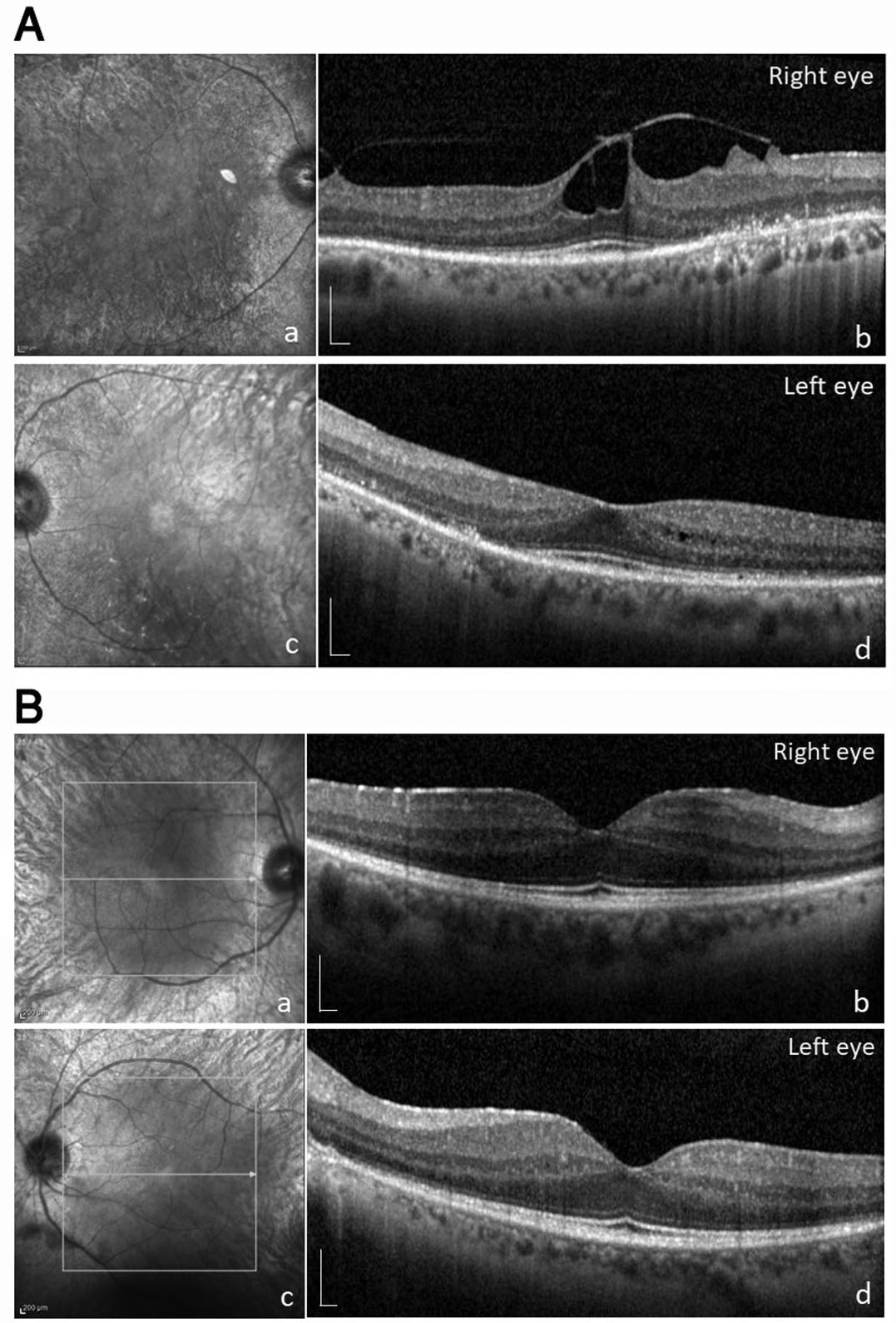
 Figure 2 of
Ali-Nasser, Mol Vis 2022; 28:359-368.
Figure 2 of
Ali-Nasser, Mol Vis 2022; 28:359-368.