Figure 3. Fundus images. A–D: Fundus photographs. E and F: Fundus autofluorescence (FAF) images. The fundus photographs of Subject 1, both A: color and B: red-free, demonstrate small drusen scattered outside and inside the vascular arcades. The images also show myelinated retinal
nerve fibers along the superior vascular arcade. C and D: Color fundus photographs of Subject 8, in whom the renewed genetic testing revealed an additional ABCA4 mutation explaining the widespread deep orange-yellow retinal flecks around the macula and in the posterior pole, typical
of Stargardt disease. E and F: The structural changes are even more evident on the FAF images, showing multiple foci of increased and reduced autofluorescence
scattered in the entire posterior pole and outside the vascular arcades.
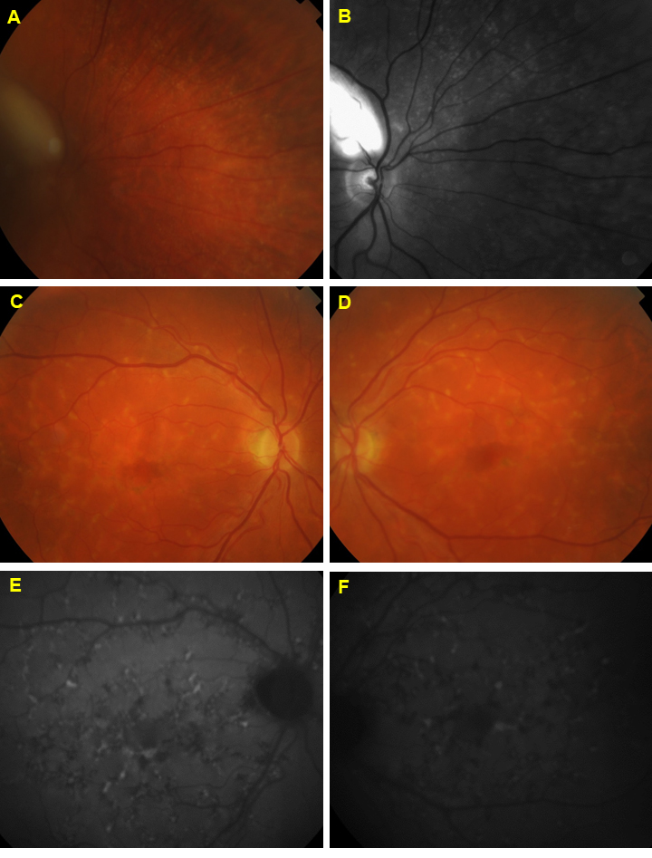
 Figure 3 of
Kjellström, Mol Vis 2022; 28:300-316.
Figure 3 of
Kjellström, Mol Vis 2022; 28:300-316.