Figure 2. Fundus images in subject 5. A: Initial and B: follow-up examination show subtle pigmentary macular changes that were quite stable over the 5-year period. The central
fundus autofluorescence (FAF) image C: at the initial examination shows a small central hyperfluorescence, which D: had slightly increased in size at the follow-up examination. The fundus photos from the E: initial and F: follow-up examinations of the right eye of Subject 7 also demonstrate stable mild pigmentary changes in the macula, whereas
the FAF images show a discrete progress of the small spots of hyperfluorescence and hypofluorescence around the vascular arcades
and in the posterior pole from G: the initial to H: the follow-up examinations.
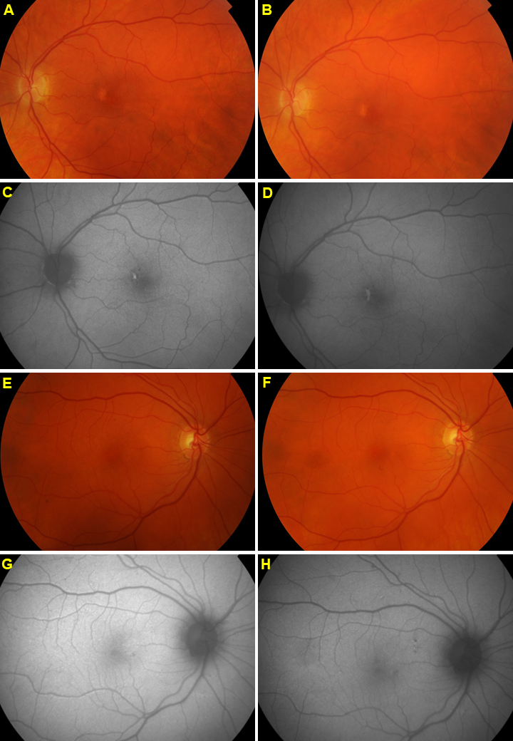
 Figure 2 of
Kjellström, Mol Vis 2022; 28:300-316.
Figure 2 of
Kjellström, Mol Vis 2022; 28:300-316.