Figure 1. Fundus images in subject 14. The fundus photographs of the right eye show quite mild macular pigmentary changes both at the
A: initial and B: follow-up examinations. The FAF images show more widespread changes with several foci of increased and reduced AF both at
the C: initial and D: follow-up examinations, and the changes worsened during the 5-year period. E: The macular OCT images confirm the changes with scarring in the RPE and interruption of the ellipsoid zone. The left eye
shows newly found structural alterations with small foci of increased and reduced AF along the vascular arcades on the FAF
image F: at the initial examination compared with G: the follow-up examination.
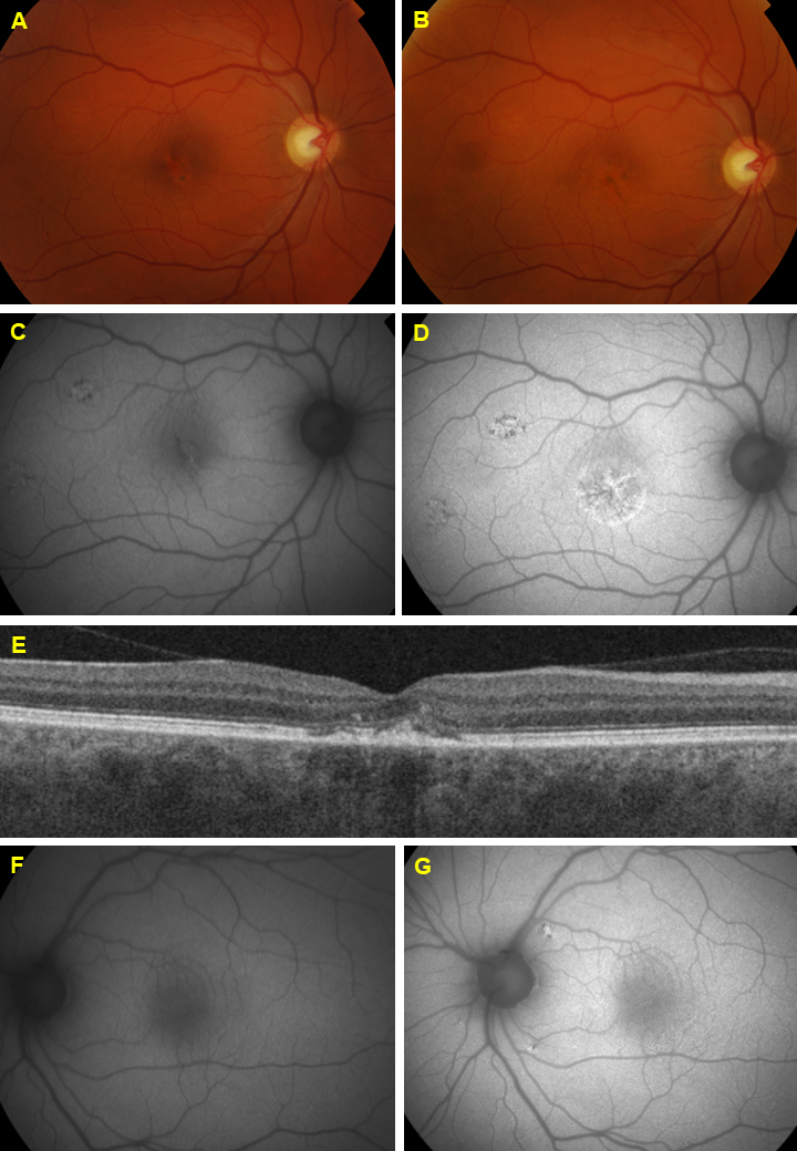
 Figure 1 of
Kjellström, Mol Vis 2022; 28:300-316.
Figure 1 of
Kjellström, Mol Vis 2022; 28:300-316.