Figure 7. Localization of Cy3 fluorescent nestin siRNAs in the retina A: with and B: without laser impact. Nestin immunoreactivity is shown in green. In A1: the retina, on the seventh day after laser impact, nestin siRNAs were abundant in some nestin-immunoreactive cells in the
GCL (rightward thick arrow) and A2: choroid (leftward thick arrow). In the control retina (retina without laser impact), nestin siRNAs were occasionally observed
in the inner retina (leftward thick arrow), B2: blood vessels in the retina (upward thick arrow), and B2: choroid (leftward thick arrow). B1: Some siRNAs appeared not integrated in the retinal cells (rightward thin arrow). Scale bars correspond to 50 μm. GCL: ganglion
cell layer, IPL: inner plexiform layer, INL: inner nuclear layer, OPL: outer plexiform layer, ONL: outer nuclear layer, Ch:
choroid.
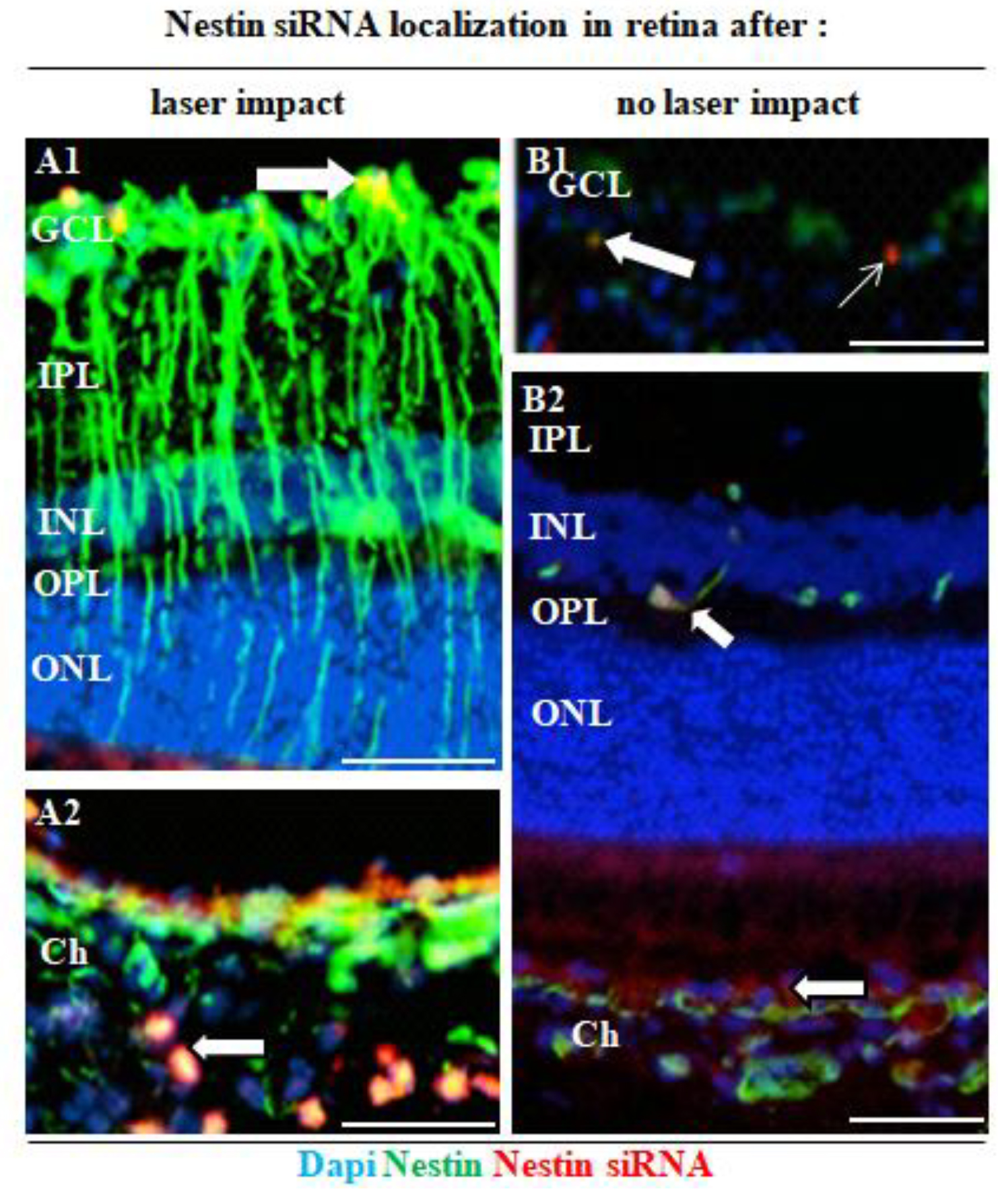
 Figure 7 of
Miloudi, Mol Vis 2022; 28:280-299.
Figure 7 of
Miloudi, Mol Vis 2022; 28:280-299.