Figure 6. von Willebrand factor (vWF, red) and nestin on the second, third, seventh, and 14th days after laser impact. A: On the second day post laser impact, vWF expression was found in blood vessels in the retina (upward thin arrow) and choroid
(downward thin arrow) at the site of the laser impact (downward thick arrow). B: Nestin immunoreactivity was found in the GCL and processes of the retina (leftward horizontal thick arrow) and choroid (rightward
horizontal thick arrow). C: Some vWF and nestin co-immunolabeling can be observed in the retina and choroid (arrowhead). D: On the third day, as in the precedent stage, vWF expression was detected in blood vessels in the retina (upward thin arrow)
and choroid (downward thin arrow) massively at the site of the laser impact (downward thick arrow). E: Nestin was detected in the retinal radial processes and blood vessels in the retina (leftward thick arrow) and choroid (rightward
thick arrow), principally at the site of the laser impact (downward thick arrow). F: vWF/nestin co-immunolabeling confirmed that blood vessels were partially immunolabeled in the retina, as in the choroid
(arrowhead). The same type of immunolabeling was found on the seventh and 14th days post laser treatment, with a progressive
decrease in the number of nestin immunolabeling (G–I and J–L). GCL: ganglion cell layer, IPL: inner plexiform layer, INL: inner nuclear layer, OPL: outer nuclear layer, ONL: outer nuclear
layer, Ch: choroid. Scale bars correspond to 50 μm.
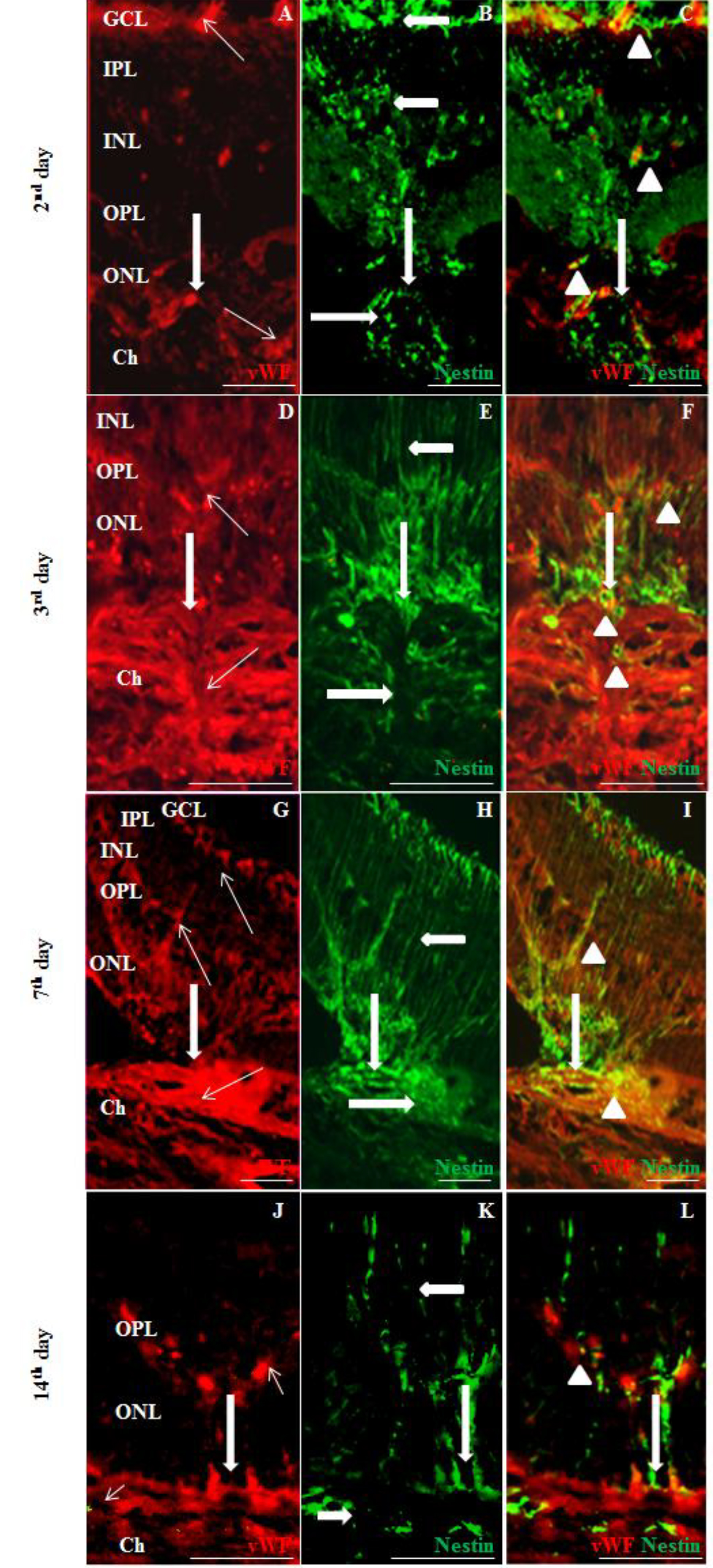
 Figure 6 of
Miloudi, Mol Vis 2022; 28:280-299.
Figure 6 of
Miloudi, Mol Vis 2022; 28:280-299.