Figure 5. Glutamine synthetase (GS; red) and nestin (green) on the second, third, seventh, and 14th days after laser impact. A: On the second day post laser treatment, GS immunoreactivity was found in functionally mature Müller cells (leftward thick
arrow) in spite of the local retinal morphology disturbance (downward thick arrow). B: Nestin immunoreactivity was observed in the GCL (upward thin arrow), as in the retinal radial processes (leftward thick
arrow), which began to enter at the choroidal level (rightward thick arrow). C: GS/nestin co-immunolabeling demonstrated no evident colocalization between the nestin and the GS. On the third day, the
laser impact was clearly detectable (downward thick arrow). D: GS was detected in Müller cells only in the retina (leftward thick arrow) despite the Brusch's membrane damage. E: Nestin immunolabeling was observed in the retinal radial processes (leftward thick arrow), clearly penetrating the choroid
(rightward thick arrow). F: GS/nestin co-immunostaining demonstrated no evident colocalization of the proteins in the retina and choroid. G: On the seventh day, as for the previous stage, GS immunolabeling was found only in Müller cells in the retina (leftward
thick arrow) despite the ruptured Bruch's membrane (downward thick arrow). Nestin immunoreactivity was observed in the radial
processes in the retina (leftward thick arrow). H: As in the precedent stage, radial processes entered into the retina (downward thick arrow) and choroid (rightward thick
arrow). I: GS/nestin co-immunolabeling demonstrated that some radial processes appeared partially co-immunolabeled in the retina (arrowhead),
but no co-immunolabeling was observed at the choroidal level (rightward thick arrow). On the 14th day, the retinal architecture
began to reform. J: GS immunolabeling is always found in Müller cells in the retina (leftward thick arrow). K: Nestin is still detected in some radial retinal processes (leftward thick arrow) and processes in the choroid (rightward
thick arrow). L: GS/nestin co-immunostaining demonstrated a colocalization of these two proteins in the retinal radial processes (arrowhead)
and confirmed the absence of GS at the choroidal level (rightward thick arrow). GCL: ganglion cell layer, IPL: inner plexiform
layer, INL: inner nuclear layer, OPL: outer nuclear layer, ONL: outer nuclear layer, Ch: choroid. Scale bars correspond to
50 μm.
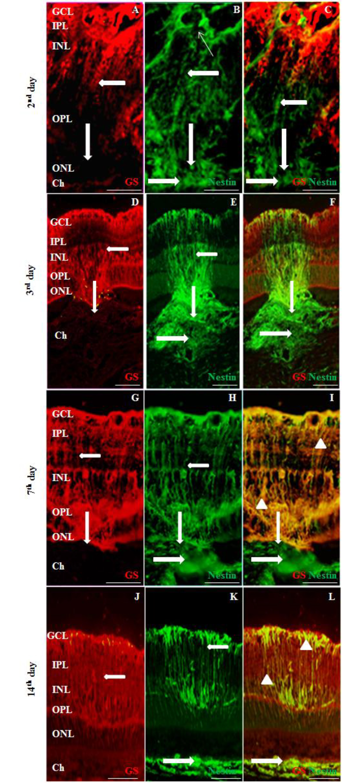
 Figure 5 of
Miloudi, Mol Vis 2022; 28:280-299.
Figure 5 of
Miloudi, Mol Vis 2022; 28:280-299.