Figure 4. Glial fibrillary acidic protein (GFAP; red) and nestin (green) on the second, third, seventh, and 14th days after laser impact.
On the second day post laser treatment, GFAP was observed mostly in the GCL (upward thin arrow) but was also found in some
retinal processes (leftward thick arrow). A: The site of the laser impact started to appear (downward thick arrow), but no processes appeared to enter the choroid. Nestin
was detected in the GCL (upward thin arrow) and retinal radial processes (leftward thick arrow). Some GFAP immunoreactive
processes also appeared at the boundary between the retina and the choroid but did not enter the choroid (rightward thick
arrow) B: Co-immunostaining confirmed these observations and allowed for the observation of a nestin/GFAP coexpression in the GCL
(upward thin arrow), C: but not in the choroid. On the third day, the laser impact was clearly detectable, along with the ruptured Bruch's membrane
(downward thick arrow). GFAP and nestin immunolabeling were observed in the D: GCL (upward thin arrow) and E: radial processes (leftward thick arrow), which clearly penetrated the choroid after the laser impact. F: GFAP/nestin co-immunostaining confirmed that some structures were co-immunolabeled in the retina and choroid (arrowhead).
On the seventh day, G: GFAP and H: nestin labeling appeared in the same structures as those in the precedent stages in the GCL (upward thin arrow) and retinal
radial processes (leftward thick arrow), but a scar began to form between the retina and the choroid (downward thick arrow),
preventing the radial retinal processes to penetrate massively in the choroid. I: Co-immunostaining confirmed the colocalization of GFAP and nestin in cells in the GCL and radial processes in the retina
(arrowhead) and a partial colocalization in the choroid (bold upper sign). On the 14th day, the morphology of the retina began
to rebuild, and a scar between the retina and the choroid became clearly visible. However, no passage of radial retinal processes
into the choroid was observed. GFAP and nestin immunolabeling are always detectable in the J: GCL (upward thin arrow) and K: retinal radial processes (leftward thick arrow), but their numbers appeared reduced. L: GFAP/nestin co-immunolabeling was observed in the GCL and retinal radial processes (arrowhead) as in the choroid (bold upper
sign). GCL: ganglion cell layer, IPL: inner plexiform layer, INL: inner nuclear layer, OPL: outer nuclear layer, ONL: outer
nuclear layer, Ch: choroid. Scale bars correspond to 50 μm.
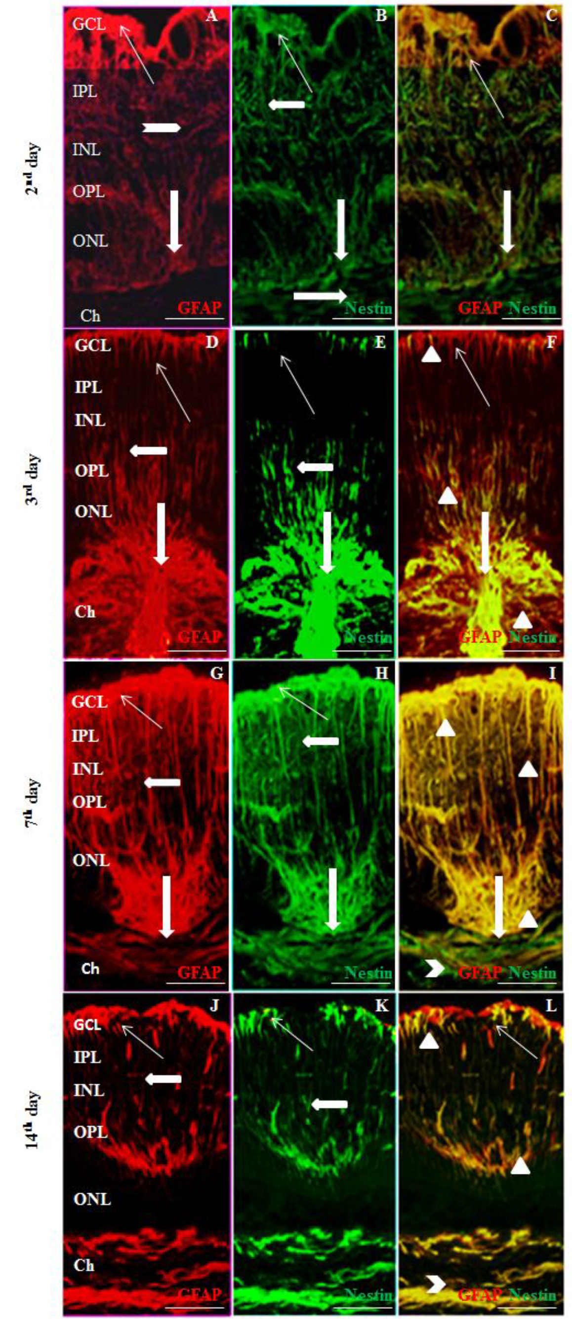
 Figure 4 of
Miloudi, Mol Vis 2022; 28:280-299.
Figure 4 of
Miloudi, Mol Vis 2022; 28:280-299.