Figure 3. Retinal and choroidal nestin (green) co-immunolabeling with glial fibrillary acidic protein (GFAP; red), glutamine synthetase
(GS; red), or von Willebrand Factor (vWF; red) at 100/150 μm from the site of the laser impact in the retina on the seventh
day. A–C: GFAP (red) and nestin (green). A: GFAP immunoreactivity is present in astrocytes (thin arrow), around blood vessels (upward thick arrow) in the GCL, in the
radial processes in the thickness of the retina (upward thin arrow), and in the choroid (arrowhead). B: Nestin is found in the same structures where GFAP is found. C: These results are confirmed by the co-immunolabeling GFAP/nestin. However, some structures (probably blood vessels) could
only be immunolabeled by nestin (downward thick arrow). D–F: GS (red) and nestin (green). D: GS immunoreactivity can be observed in Müller cells (thin arrow) and sometimes at the choroidal level (arrowhead). E: Nestin is found in in the GCL (upward thin arrow), in the radial processes in the thickness of the retina (downward thin
arrow), and in the choroid (arrowhead). F: Partial GS/nestin co-immunolabeling is rarely and partially observed in the radial processes (downward thin arrow) and GCL
(upward thin arrow). No co-immunolabeling was found in the choroid (arrowhead) and probably in blood vessels at the choroidal
level (arrowhead). G–I: vWF (red) and nestin (green). G: vWF can be observed in blood vessels throughout the retina (upward thin arrow) and choroid (horizontal thin arrow). H: Nestin was found in cells and processes in the retina (upward thin arrow). It was also detected in blood vessels in the
retina (downward thin arrow) and choroid. I: Co-immunolabeling confirmed the presence of nestin and vWF in some blood vessels in the retina (thick arrow) and choroid.
Scale bars correspond to 50 μm. GCL: ganglion cell layer, IPL: inner plexiform layer, INL: inner nuclear layer, OPL: outer
plexiform layer, ONL: outer nuclear layer, Ch: choroid.
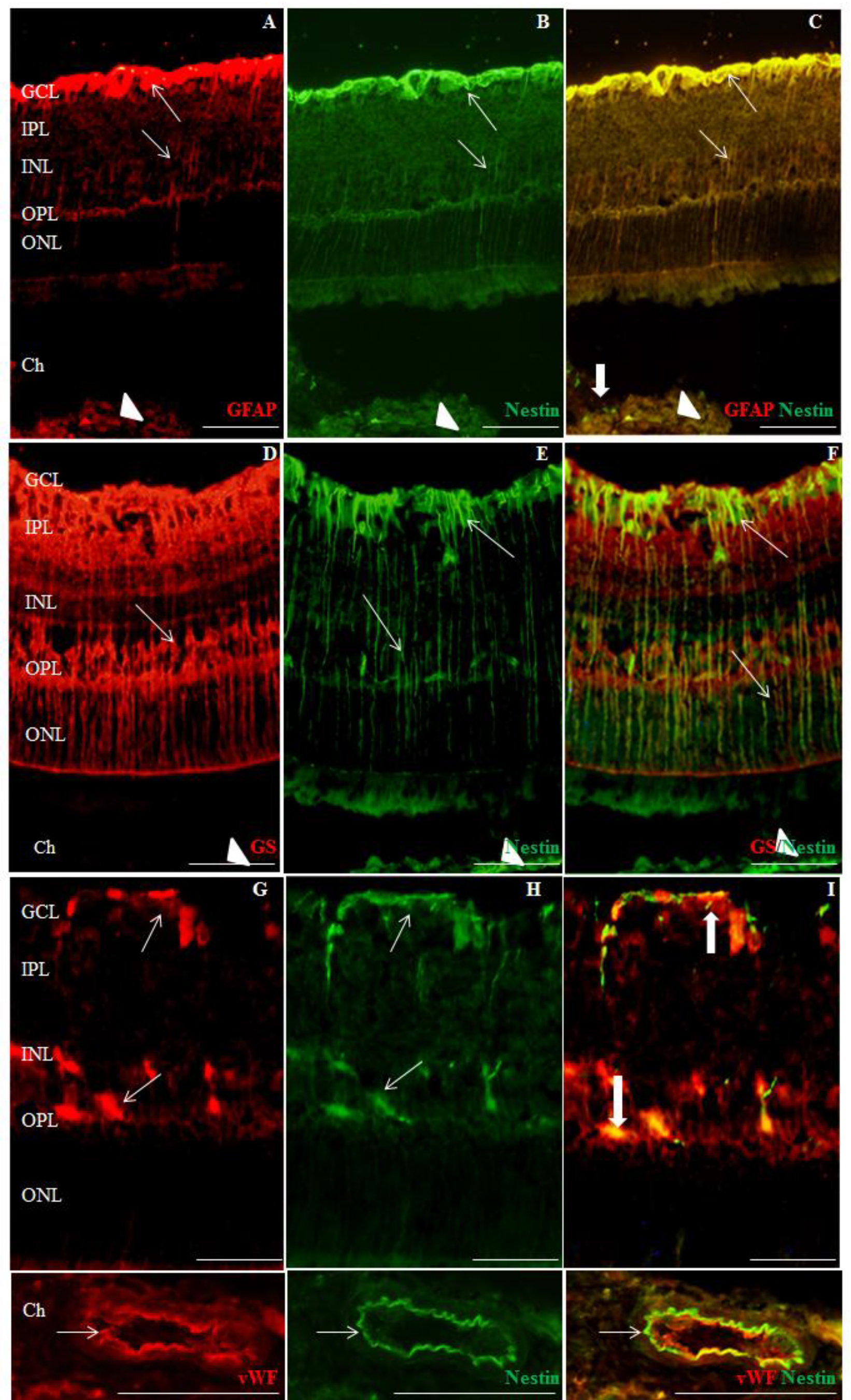
 Figure 3 of
Miloudi, Mol Vis 2022; 28:280-299.
Figure 3 of
Miloudi, Mol Vis 2022; 28:280-299.