Figure 1. Histological pictures of the retina and choroid in the rats not treated with krypton laser impact, at a distance of 150 μm
from the site where laser impact was to be applied and at the site of impact. Immunofluorescent labeling with DAPI (blue)
and nestin (green) is shown. A, C, G, I, M, O, S, and U: No morphological changes can be observed in the control rats not treated with laser impact and at 150 μm from the site of
laser impact, as observed with DAPI staining. Nestin immunostaining can be seen in the processes of astrocytes and in the
probable end feet of Müller cells located mostly in the GCL (thin arrow). B, D, H, J, N, P, T, and V: Blood vessels are also located in the retina and choroid (horizontal large arrow). E, K, Q, and W: At the impact, DAPI labeling demonstrated that retinal morphology is greatly disrupted in the outer retina at all stages
examined, with cells of the ONL entering the choroid (large vertical thick arrow), especially evident on the second and third
days after laser impact. F: Nestin immunostaining was observed from the second day in glial cells in the GCL (thin arrow), in the retinal radial processes
(horizontal large arrow), and in the choroidal processes (large star). The immunolabeling remained intense on the L: third day and R: seventh day, when an edema appeared to start to form (bold upper sign), but was decreased on the X: 14th day. Scale bars correspond to 50 μm. All pictures were taken from different retinal sections. GCL: ganglion cell layer,
IPL: inner plexiform layer, INL: inner nuclear layer, OPL: outer plexiform layer, ONL: outer nuclear layer, Ch: choroid.
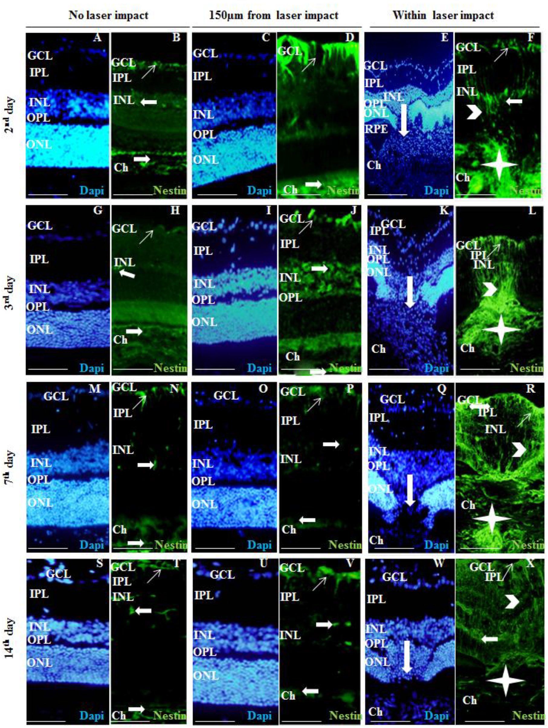
 Figure 1 of
Miloudi, Mol Vis 2022; 28:280-299.
Figure 1 of
Miloudi, Mol Vis 2022; 28:280-299.