Figure 2. Clinical photography. The slit lamp imaging shows A: iris transillumination defects, B: a persistent pupillary membrane, and C: posterior lentiglobus or spherophakia on the slit illumination observed in Individual A-II-2. D: Ectopia lentis seen in Individual A-II-1 OD.
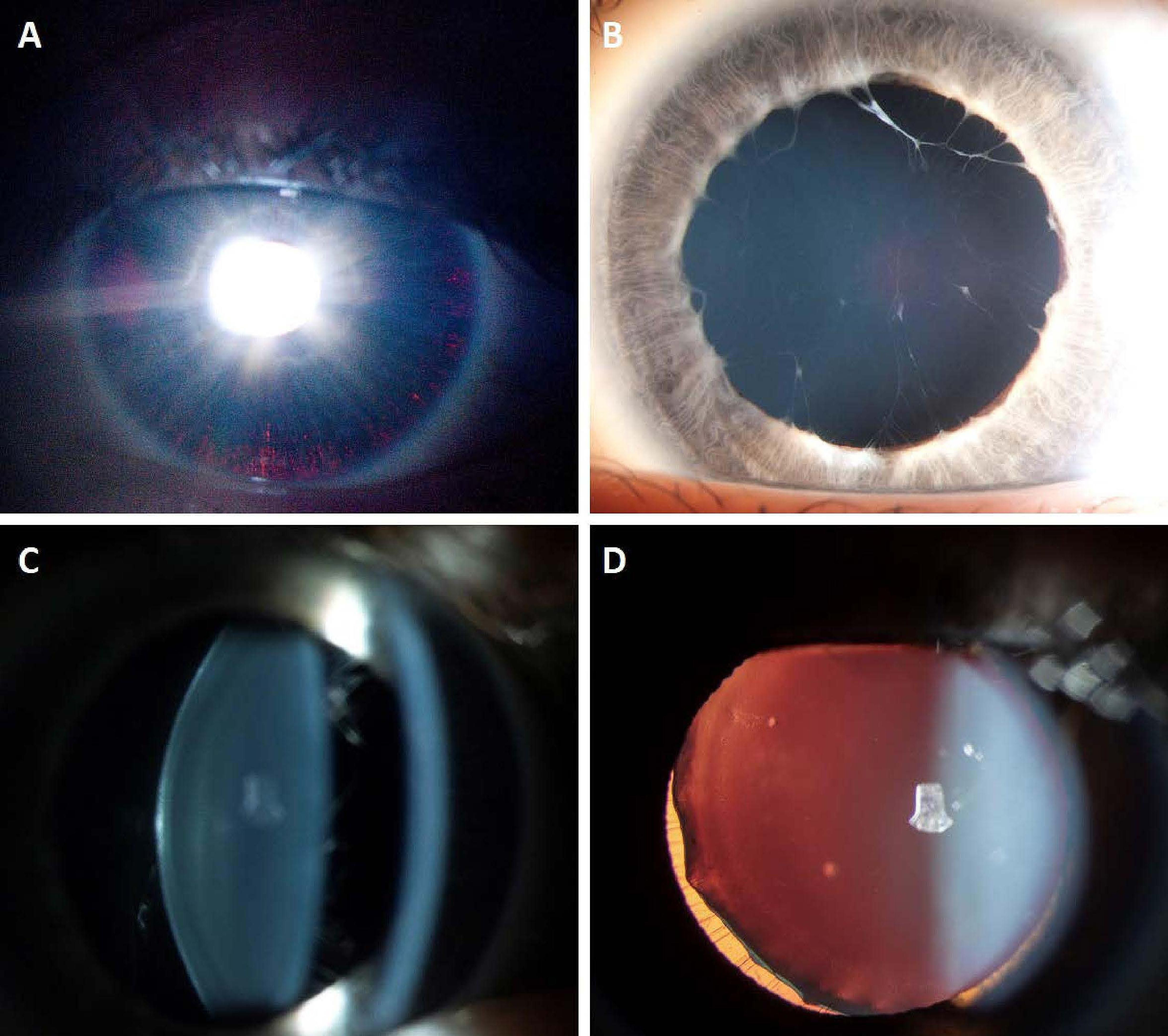
 Figure 2 of
Knight, Mol Vis 2022; 28:257-268.
Figure 2 of
Knight, Mol Vis 2022; 28:257-268.