Figure 1. Removal of extracellular calcium induced Lucifer yellow (LY) uptake in lens fiber cells, which was correlated with ATP release.
A, B: Time course of ATP release into the media measured from rat lenses incubated in either AAH (black) or 0 mM [Ca2+]e AAH (blue). The error bars represent the standard error of the mean, and “()” represents the number of lenses in each experimental
group. **p <0.01. C-F: Images of Texas red-dextran (TRD; C, E) and LY (D, F) penetration in equatorial sections from lenses incubated for 1 h in either the control AAH (C, D) or the 0 mM [Ca2+]e AAH (E, F). G: Normalized LY signal intensity plotted against distance into the lens taken from the lenses incubated in AAH (green line) or 0 mM [Ca2+]e AAH (dashed green line) showing an area (***) of LY uptake induced by 0 mM [Ca2+]e. (H) Normalized LY (dashed green line) and TRD (red line) signal intensities plotted against distance into the lens taken
from the lenses incubated in 0 mM [Ca2+]e AAH. Scale bar = 50 µm.
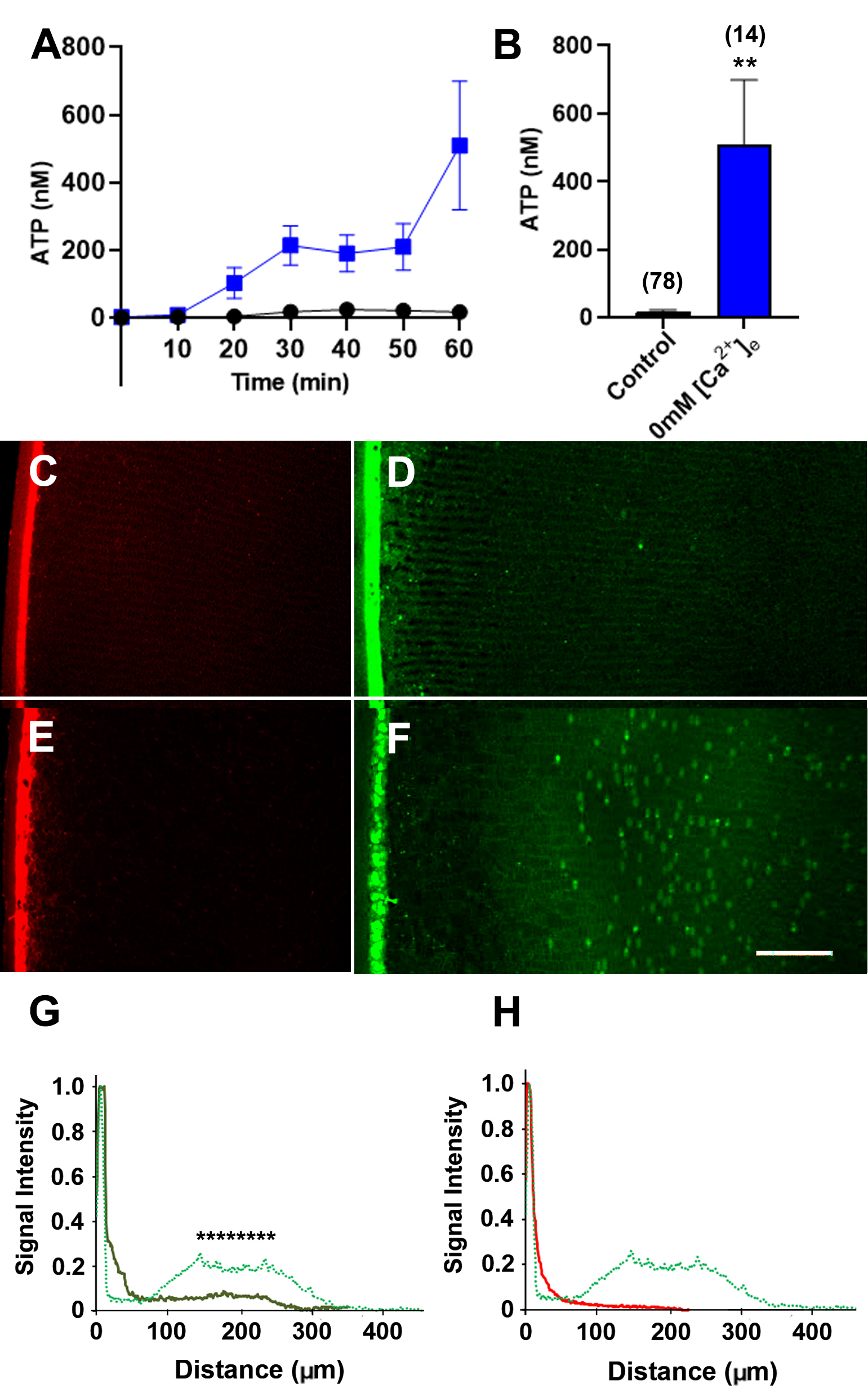
 Figure 1 of
Suzuki-Kerr, Mol Vis 2022; 28:245-256.
Figure 1 of
Suzuki-Kerr, Mol Vis 2022; 28:245-256.