Figure 1. Optical coherence tomography angiography (OCT-A) and segmentation. A: 6 × 6 mm OCT-A scan of the whole macula superficial vascular complex (SVC) area (1). Contained within the whole area used
to calculate the whole VD, including the fovea (2) and parafovea (3–6). B: The B scan indicates the automatic separation of the SVC from the DVC at the inner plexiform layer (IPL)/inner nuclear layer
(INL) segmented line in blue. The SVC is defined from the vitreous/ILM segmented line (red) to the IPL/INL segmented line
(green), while the deep vascular complex (DVC) is defined from the IPL/INL segmented line (green) to the OPL/ONL segmented
line (blue). The VD of the SVC (D) and the scanning light ophthalmoscopy (E) are shown for reference.
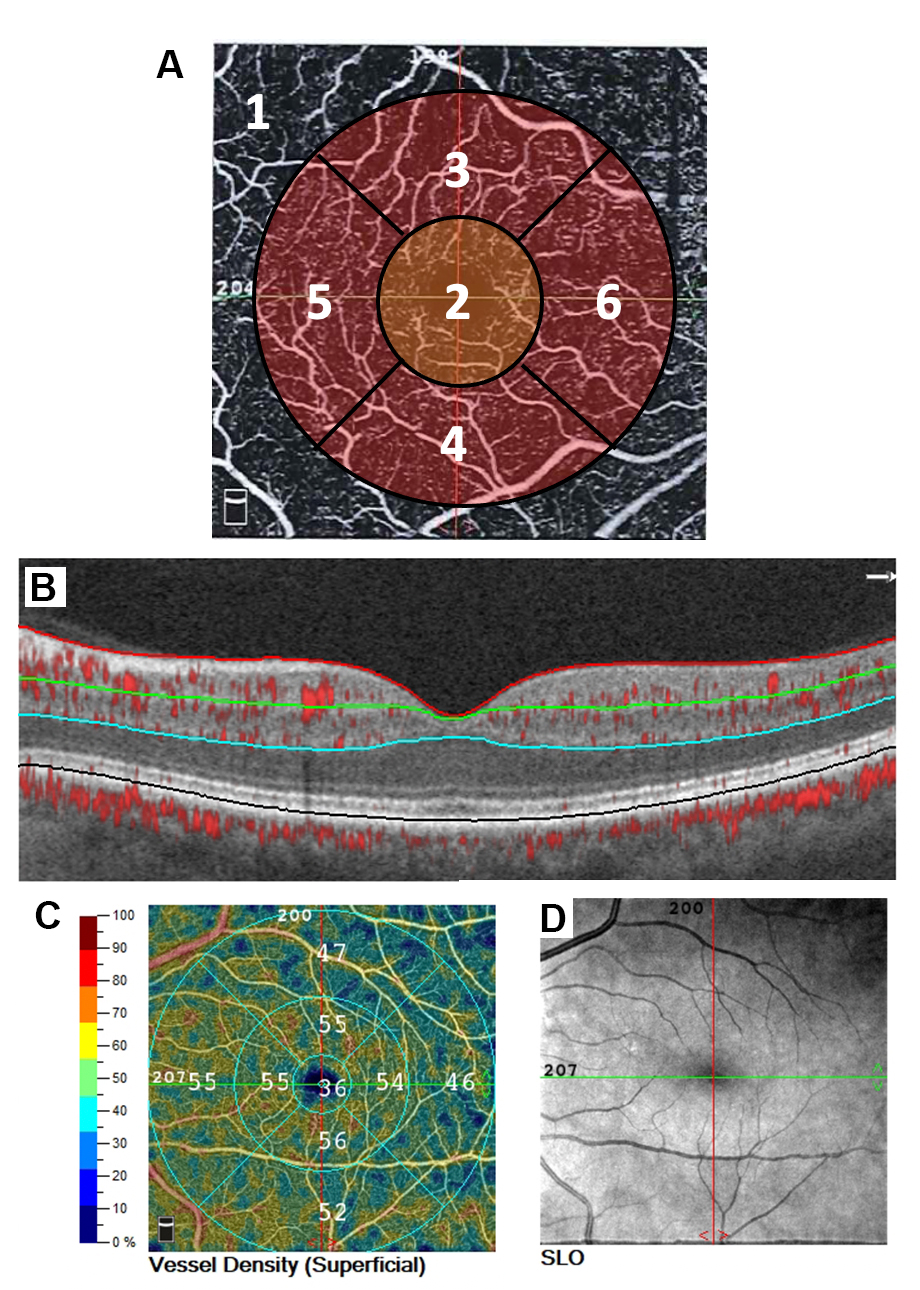
 Figure 1 of
Scheive, Mol Vis 2022; 28:220-229.
Figure 1 of
Scheive, Mol Vis 2022; 28:220-229.