Figure 1. Color fundus, fundus autofluorescence (FAF), and spectral-domain optical coherence tomography (SD-OCT) images of the patients
with exon 3 haplotype blue cone monochromacy. (A–F) MOL0057–3, (G–L) MOL1383–1, (M–R) MOL1434–1, and (S–X) MOL1736–1. (A, B, G, H, M, N,S, and T) Color fundus photos showing peripapillary atrophy, temporal pallor of the optic disc, normal-looking peripheral retina,
and disrupted foveal reflex, except MOL1383–1. The parallel FAF images (C, D, O, P,U, and V) demonstrate a hyperfluorescent foveal reflex complementary with foveal ellipsoid zone atrophy in the SD-OCT horizontal cross-sections
(E, F, Q, R,W, and X). (I, J) Normal FAF reflex for MOL1383–1 reflecting the preserved ellipsoid zone in the SD-OCT cross-sectional images (K, L).
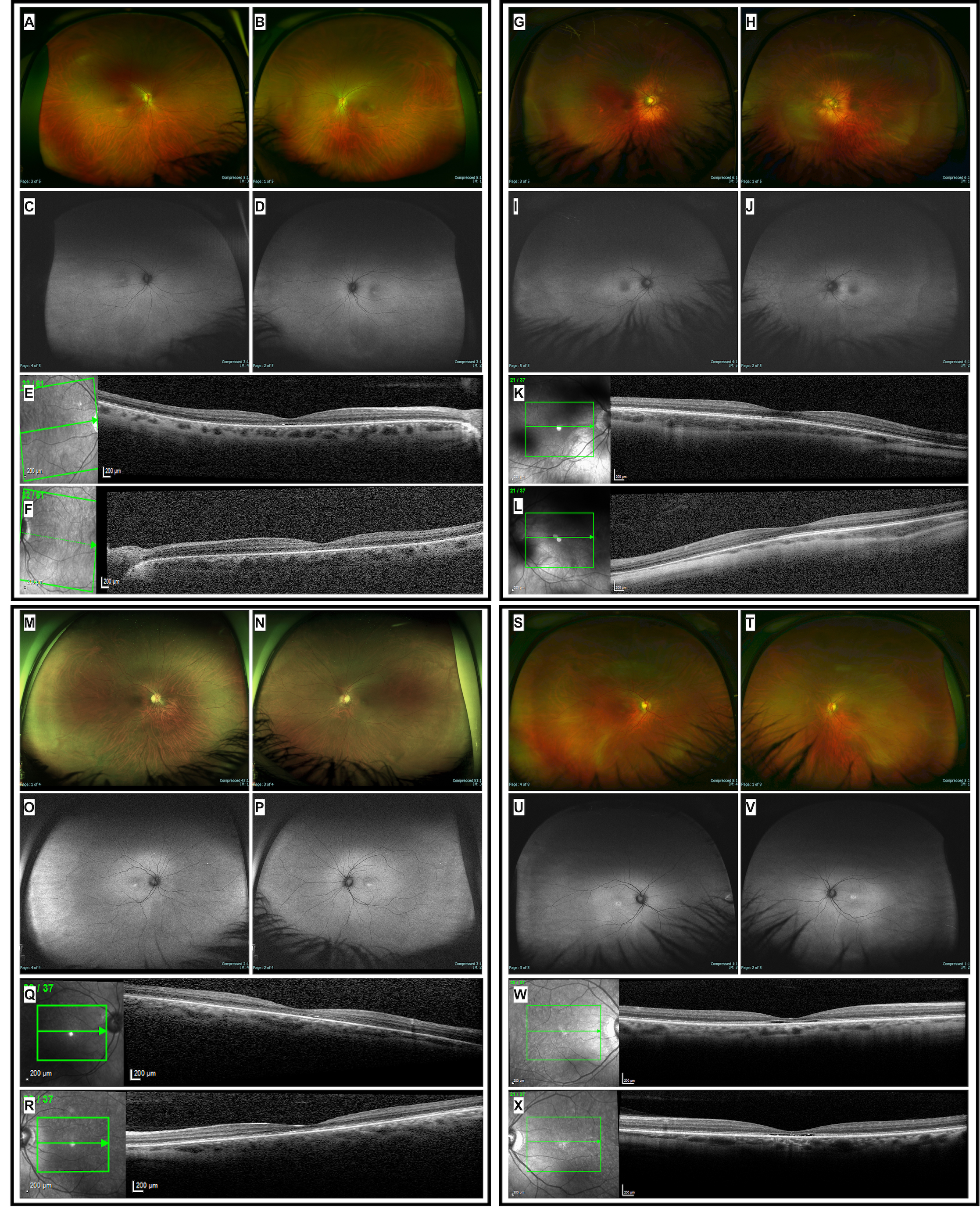
 Figure 1 of
Khateb, Mol Vis 2022; 28:21-28.
Figure 1 of
Khateb, Mol Vis 2022; 28:21-28.