Figure 5. PT35 had a pseudo-vitelliform clinical phenotype compatible with occult macular dystrophy (OMD) and found to harbor a novel
RP1L1 change. A: The macular OCT image of the right eye, which was symptomatic of metamorphopsia, demonstrates a dome-shaped pocket of subfoveal
hyporeflective fluid with a fragmented subretinal hyperreflective material (green arrow). B: The macular OCT image of the left eye, which was asymptomatic at presentation, shows a tiny pseudovitelliform subfoveal
hyperreflective lesion (white arrow). C: Unlike the prediction from these focal findings, which were minimal in the left eye, the response densities of the multifocal
electroretinogram were markedly depressed in both eyes, with partial foveal peak preservation, a common finding in OMD. The
BEST1 gene sequencing was normal, as was the EOG (not shown).
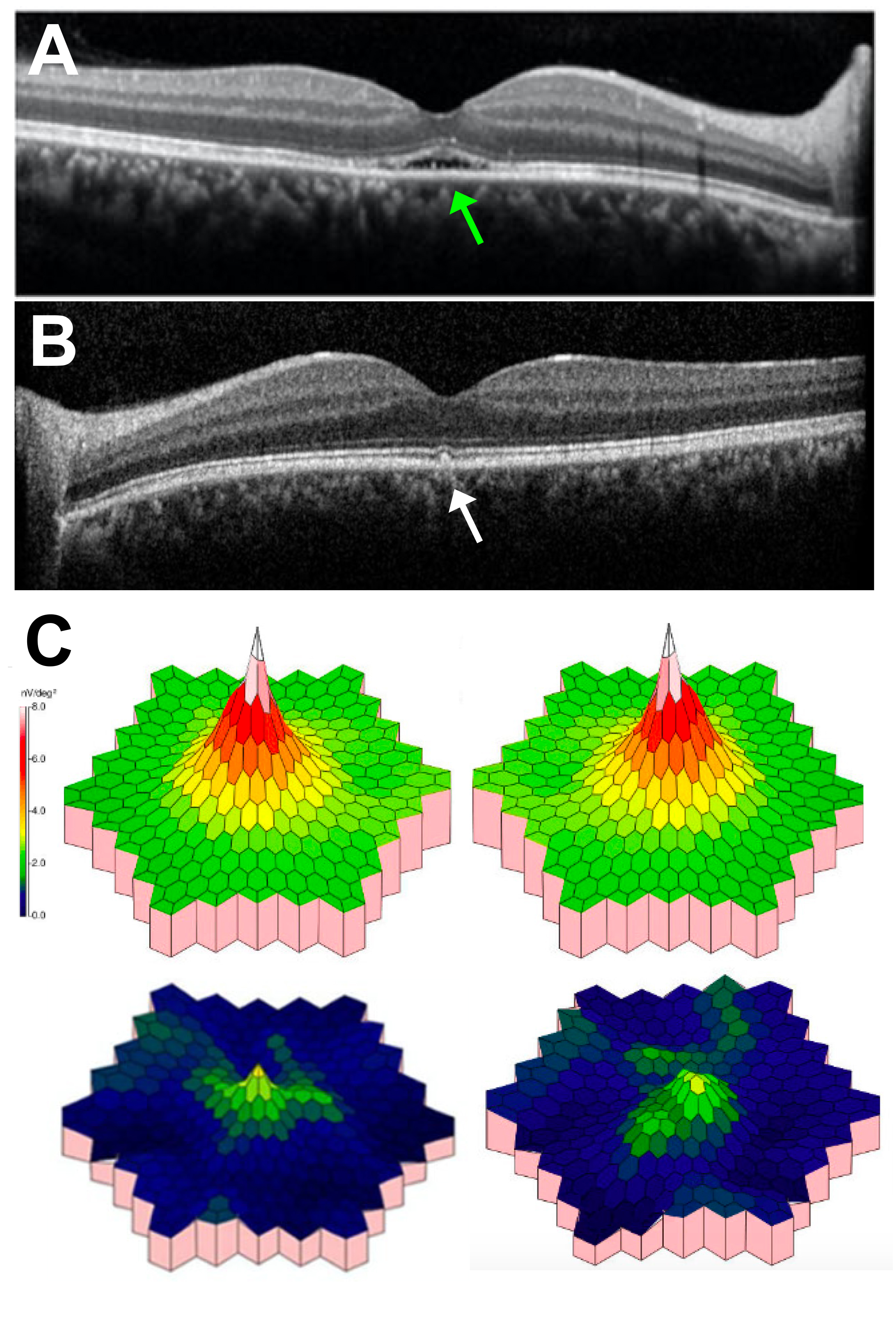
 Figure 5 of
Gupta, Mol Vis 2022; 28:203-219.
Figure 5 of
Gupta, Mol Vis 2022; 28:203-219.